Review
. 2012 Oct;37(10):425-35.
doi: 10.1016/j.tibs.2012.06.006. Epub 2012 Jul 23.

Structural dynamics of nucleosomes at single-molecule resolution

Affiliations
Review

Structural dynamics of nucleosomes at single-molecule resolution

John S Choy et al. Trends Biochem Sci. 2012 Oct.

Abstract

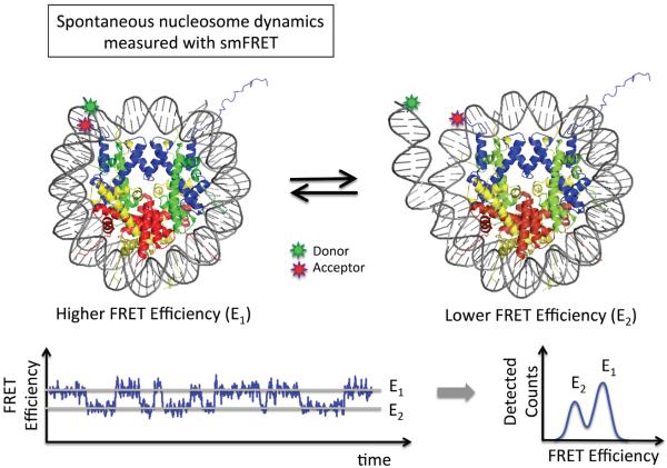
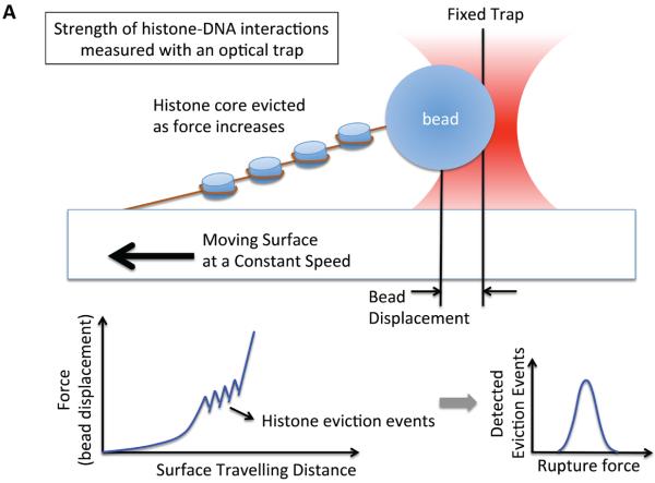
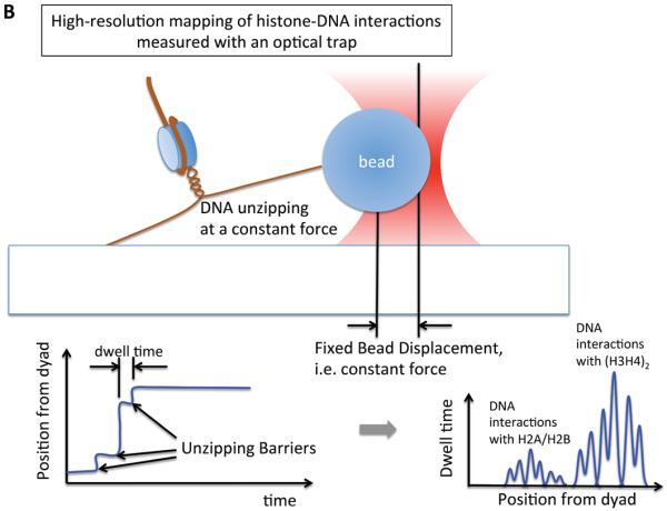
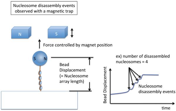
The detailed mechanisms of how DNA that is assembled around a histone core can be accessed by DNA-binding proteins for transcription, replication, or repair, remain elusive nearly 40 years after Kornberg's nucleosome model was proposed. Uncovering the structural dynamics of nucleosomes is a crucial step in elucidating the mechanisms regulating genome accessibility. This requires the deconvolution of multiple structural states within an ensemble. Recent advances in single-molecule methods enable unprecedented efficiency in examining subpopulation dynamics. In this review, we summarize studies of nucleosome structure and dynamics from single-molecule approaches and how they advance our understanding of the mechanisms that govern DNA transactions.


Figures

Figure 1
Figure 1. Single molecule fluorescence resonance energy transfer (FRET) studies on the structure and structural dynamics of nucleosomes
FRET labels are attached to DNA and/or histones to elucidate the structure of nucleosomes and to follow the dynamic unwrapping and/or rewrapping of the nucleosomal DNA. The fluorophore positions in the example shown are from the fluorescence correlation spectroscopy (FCS) study by the Widom group [22]. The time trajectory of FRET efficiency or FCS measurement reveals dynamic motions of the nucleosomal DNA termini within the temporal resolution of the measurement. A simulated ideal FRET time trajectory of a surface immobilized single nucleosome particle is shown to illustrate the measurement scheme. The time-averaged FRET efficiency histogram reveals the closed (E1; high FRET) and opened (E2; low FRET) states of a nucleosome.
Figure 2
Figure 2. Single molecule studies of nucleosomes with optical tweezers
(a) A setup based on using optical trapping to study the interaction strengths between DNA and histones in a nucleosome array. One end of the array is attached to a glass surface and the other end is attached to a plastic bead that is trapped within a focus of a laser beam. By moving the slide in one direction, the force to elongate the array is gradually increased. As the force increases, histones are evicted and the force (i.e. the distance between the two points of attachment) decreases sharply at the moment of eviction because histone eviction yields increases in the length of the array. Multiple histone eviction events are observed within an array as depicted in the chart. (b) A setup based on optical trapping that gives a high-resolution map of interactions between nucleosomal DNA and core histones [31]. Each end of the nucleosomal DNA is anchored and the measurements were made for a single nucleosome. The force to unzip the DNA is held constant by maintaining the distance between the two anchored points constant. When the applied force is slightly above the unzipping force, DNA is unzipped stepwise through multiple unzipping barriers. When the resolution of force control/measurement is high, the unzipping barriers would be detected as schematized in the chart and bear information on the interactions between the DNA and histones within a nucleosome.
Figure 3
Figure 3. Single molecule studies of nucleosome structure with magnetic tweezers
A magnetic tweezers setup to observe nucleosome eviction events in a nucleosome array. One end of the array is attached to a glass surface and the other end is attached to a magnetic bead trapped in a magnetic field. The vertical position of the magnet is held constant to apply a constant force along the array. When the force is slightly above the nucleosome rupture force, multiple nucleosome dissociation events are detected as depicted in the chart.
Figure 4
Figure 4. Single molecule studies of ATP-dependent chromatin remodeling
(a) A single molecule fluorescence resonance energy transfer (FRET) setup to study nucleosome sliding by the ACF remodeling complex [74]. Three FRET populations were observed from nucleosomes: one from D1A FRET pair (the peak marked as D1A in the FRET histogram), another from D2A pair (peak D2A in the histogram) and the third from the two-donor and one-acceptor FRET (some nucleosomes have two donor fluorophores according to the stoichiometry of the labeled histone in the histone core, marked D1A+D2A in the histogram). Consistent with nucleosome sliding mediated by ACF, a dramatic FRET change was observed upon the addition of ACF and ATP. (b) An optical tweezers setup to monitor the structure of a short nucleosome array altered by SWI/SNF or RSC remodeling complexes in the presence of ATP [75]. Several reversible changes in the length of the array were observed upon the addition of SWI/SNF (or RSC) and ATP, suggesting DNA looping events induced by SWI/SNF remodeling. (c) A magnetic tweezers setup used to study the topology change of DNA caused by the remodeling activity of the RSC complex [77]. The study revealed that RSC induces superhelical loops of DNA in an ATP dependent manner.

Similar articles

Cited by

References

    1. Kornberg RD. Chromatin structure: a repeating unit of histones and DNA. Science. 1974;184:868–871. - PubMed
    1. Kornberg RD, Thomas JO. Chromatin structure; oligomers of the histones. Science. 1974;184:865–868. - PubMed
    1. Kornberg RD. Structure of chromatin. Annu Rev Biochem. 1977;46:931–954. - PubMed
    1. Richmond TJ, et al. Structure of the nucleosome core particle at 7 A resolution. Nature. 1984;311:532–537. - PubMed
    1. Klug A, et al. A low resolution structure for the histone core of the nucleosome. Nature. 1980;287:509–516. - PubMed

Publication types

LinkOut - more resources