

A novel distinctive cerebrovascular phenotype is associated with heterozygous Arg179 ACTA2 mutations
- PMID: 22831780
- PMCID: PMC3407424
- DOI: 10.1093/brain/aws172
A novel distinctive cerebrovascular phenotype is associated with heterozygous Arg179 ACTA2 mutations
Abstract
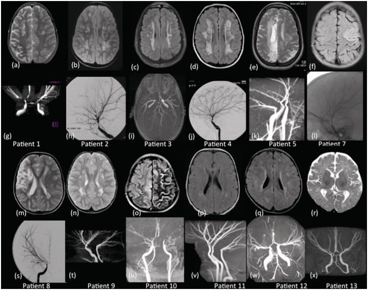
Mutations in the ACTA2 gene lead to diffuse and diverse vascular diseases; the Arg179His mutation is associated with an early onset severe phenotype due to global smooth muscle dysfunction. Cerebrovascular disease associated with ACTA2 mutations has been likened to moyamoya disease, but appears to have distinctive features. This study involved the analysis of neuroimaging of 13 patients with heterozygous missense mutations in ACTA2 disrupting Arg179. All patients had persistent ductus arteriosus and congenital mydriasis, and variable presentation of pulmonary hypertension, bladder and gastrointestinal problems associated with this mutation. Distinctive cerebrovascular features were dilatation of proximal internal carotid artery, occlusive disease of terminal internal carotid artery, an abnormally straight course of intracranial arteries, and absent basal 'moyamoya' collaterals. Patterns of brain injury supported both large and small vessel disease. Key differences from moyamoya disease were more widespread arteriopathy, the combination of arterial ectasia and stenosis and, importantly, absence of the typical basal 'moyamoya' collaterals. Evaluation of previously published cases suggests some of these features are also seen in the ACTA2 mutations disrupting Arg258. The observation that transition from dilated to normal/stenotic arterial calibre coincides with where the internal carotid artery changes from an elastic to muscular artery supports the hypothesis that abnormal smooth muscle cell proliferation caused by ACTA2 mutations is modulated by arterial wall components. Patients with persistent ductus arteriosus or congenital mydriasis with a label of 'moyamoya' should be re-evaluated to ensure the distinctive neuroimaging features of an ACTA2 mutation have not been overlooked. This diagnosis has prognostic and genetic implications, and mandates surveillance of other organ systems, in particular the aorta, to prevent life-threatening aortic dissection.
Figures
References
-
- Ades LC, Davies R, Haan EA, Holman KJ, Watson KC, Sreetharan D, et al. Aortic dissection, patent ductus arteriosus, iris hypoplasia and brachytelephalangy in a male adolescent. Clin Dysmorphol. 1999;8:269–76. - PubMed
-
- Disabella E, Grasso M, Gambarin FI, Narula N, Dore R, Favalli V, et al. Risk of dissection in thoracic aneurysms associated with mutations of smooth muscle alpha-actin 2 (ACTA2) Heart. 2011;97:321–6. - PubMed
-
- Fukui M. Guidelines for the diagnosis and treatment of spontaneous occlusion of the circle of Willis (‘moyamoya’ disease). Research Committee on Spontaneous Occlusion of the Circle of Willis (Moyamoya Disease) of the Ministry of Health and Welfare, Japan. Clin Neurol Neurosurg. 1997;99(Suppl 2):S238–40. - PubMed
-
- Fullerton HJ, Wu YW, Sidney S, Johnston SC. Risk of recurrent childhood arterial ischemic stroke in a population-based cohort: the importance of cerebrovascular imaging. Pediatrics. 2007;119:495–501. - PubMed
Publication types
MeSH terms
Substances
Grants and funding
LinkOut - more resources
Full Text Sources
Miscellaneous
