. 2012 Jan 26;1(1):2-12.
doi: 10.1016/j.celrep.2011.11.001. Epub 2011 Dec 15.

Mutations in the gene PRRT2 cause paroxysmal kinesigenic dyskinesia with infantile convulsions

Affiliations

Mutations in the gene PRRT2 cause paroxysmal kinesigenic dyskinesia with infantile convulsions

Hsien-Yang Lee et al. Cell Rep. .

Abstract

Paroxysmal kinesigenic dyskinesia with infantile convulsions (PKD/IC) is an episodic movement disorder with autosomal-dominant inheritance and high penetrance, but the causative genetic mutation is unknown. We have now identified four truncating mutations involving the gene PRRT2 in the vast majority (24/25) of well-characterized families with PKD/IC. PRRT2 truncating mutations were also detected in 28 of 78 additional families. PRRT2 encodes a proline-rich transmembrane protein of unknown function that has been reported to interact with the t-SNARE, SNAP25. PRRT2 localizes to axons but not to dendritic processes in primary neuronal culture, and mutants associated with PKD/IC lead to dramatically reduced PRRT2 levels, leading ultimately to neuronal hyperexcitability that manifests in vivo as PKD/IC.


Figures

Figure 1
Figure 1. Twenty-four of twenty-five PKD/IC pedigrees with most secure phenotypes carry PRRT2 mutations
Eleven of these 24 PKD/IC pedigrees are shown. Females are denoted with circles and males with squares. The kindred number is denoted at the upper left corner of each pedigree and the DNA number are noted under individuals where it was available. The specific mutation is denoted under each individual, when present. Individuals with a DNA number but no mutation noted have the wild-type genotype. Affection status for PKD, Infantile Convulsions (under age 2 years), and GS (generalized seizures occurring after age 2 years) are as noted. Samples that were used for WGS are marked with an asterisk (*). One phenocopy was present in K3323 (marked by an arrow).
Figure 2
Figure 2. Figure 1: Twenty-four of twenty-five PKD/IC pedigrees with most secure phenotypes carry PRRT2 mutations
Thirteen of these PKD/IC pedigrees are shown. Symbols are as in Figure 1.
Figure 3
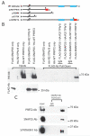
Figure 3. PRRT2 and SNAP25 interact in vitro and in vivo
(A) The comparison of protein structures for WT and truncated mutants of PRRT2. The blue rectangles represent putative C-terminal transmembrane domains of PRRT2. The black arrows represent positions of mutations producing either nonsense or frameshift mutations. Red rectangles represent novel protein sequences produced by frame shift mutations. (B) In vitro co-immunoprecipitation was performed in HEK293T cells singly transfected, or co-transfected with FLAG-tagged SNAP25 and either HA-tagged WT or mutant forms (p.R217Pfs*8) of PRRT2. After FLAG antibody pull-down, only the cell extract from HEK293T cells co-transfected with FLAG-tagged SNAP25 and HA-tagged WT PRRT2 showed an ~65 kDa band (upper panel, right-most lane), implying an interaction exists between SNAP25 and PRRT2 in vitro. Interestingly, no obvious expression was detected in cell extracts transfected with the mutant form of PRRT2 (upper panel, the second and the fourth lanes from the left). (C) In vivo co-immunoprecipitation of Snap25 and Prrt2 using whole brain extracts from a control mouse. After SNAP25 antibody pull-down, a Prrt2 band (~65 kDa) was detected using anti-PRRT2 antibody. An antibody specific for SYNTAXIN1, a protein interacting with SNAP25, was also used as a positive control.
Figure 4
Figure 4. Truncated mutations of PRRT2 lead to abnormal protein expression in vitro
When HEK293T cells were co-transfected with FLAG-tagged PRRT2 and HA-tagged PRRT2, an ~65 kDa band was present when probed with antibody for the tag on the WT fusion protein but FLAG-tagged fusion proteins for the truncation mutations showed a significant reduction (R240X and I327Ifs*14) or undetectable expression (R217pfs*8 and E173X). Thus, the mutations led to low or undetectable PRRT2 protein levels that did not affect the wild type allele in vitro. GAPDH antibody was used as a sample loading control.
Figure 5
Figure 5. Expression and localization of PRRT2 in hippocampal neurons
(A) Co-immunostaining of WT FLAG-PRRT2 and MAP2 showed distinct localization patterns. (B) Co-immunostaining for WT FLAG-PRRT2 and synapsin I showed WT PRRT2 co-localized with synapsin I in neuronal puncta. (C) When co-immunostaining of the FLAG-PRRT2 R217Pfs*8 mutant with synapsin I, no obvious positive staining of mutant PRRT2 was detected. Red= WT FLAG-PRRT2; Green=MAP2 or Synapsin1. Scale bars=10 microns.

References

    1. Abe T, Kobayashi M, Araki K, Kodama H, Fujita Y, Shinozaki T, Ushijima H. Infantile convulsions with mild gastroenteritis. Brain & development. 2000;22:301–306. - PubMed
    1. Bennett LB, Roach ES, Bowcock AM. A locus for paroxysmal kinesigenic dyskinesia maps to human chromosome 16. Neurology. 2000;54:125–130. - PubMed
    1. Bhatia KP. Paroxysmal dyskinesias. Mov Disord. 2011;26:1157–1165. - PubMed
    1. Blakeley J, Jankovic J. Secondary paroxysmal dyskinesias. Mov Disord. 2002;17:726–734. - PubMed
    1. Bruno MK, Hallett M, Gwinn-Hardy K, Sorensen B, Considine E, Tucker S, Lynch DR, Mathews KD, Swoboda KJ, Harris J, et al. Clinical evaluation of idiopathic paroxysmal kinesigenic dyskinesia: new diagnostic criteria. Neurology. 2004;63:2280–2287. - PubMed

Publication types

MeSH terms

Supplementary concepts