. 2012 Jan 26;1(1):13-20.
doi: 10.1016/j.celrep.2011.11.004. Epub 2011 Dec 15.

Premetazoan origin of the hippo signaling pathway

Affiliations

Premetazoan origin of the hippo signaling pathway

Arnau Sebé-Pedrós et al. Cell Rep. .

Abstract

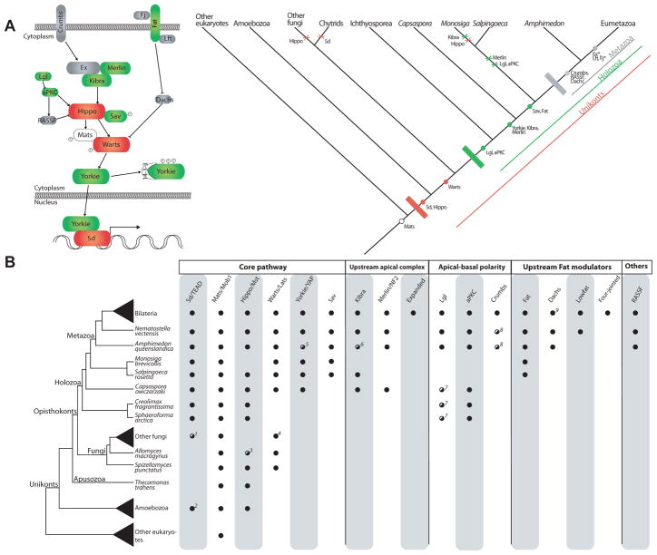
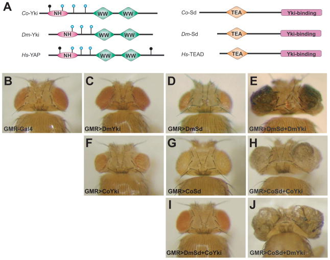
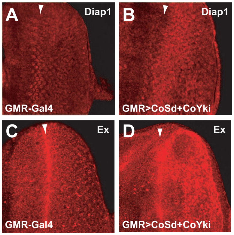
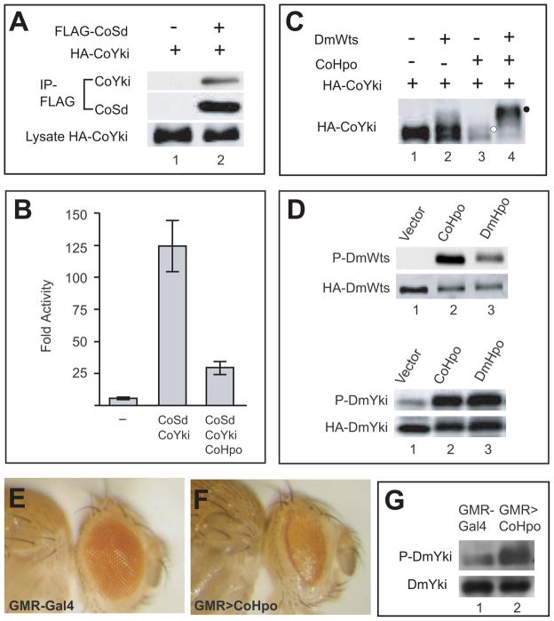
Nonaggregative multicellularity requires strict control of cell number. The Hippo signaling pathway coordinates cell proliferation and apoptosis and is a central regulator of organ size in animals. Recent studies have shown the presence of key members of the Hippo pathway in nonbilaterian animals, but failed to identify this pathway outside Metazoa. Through comparative analyses of recently sequenced holozoan genomes, we show that Hippo pathway components, such as the kinases Hippo and Warts, the coactivator Yorkie, and the transcription factor Scalloped, were already present in the unicellular ancestors of animals. Remarkably, functional analysis of Hippo components of the amoeboid holozoan Capsaspora owczarzaki, performed in Drosophila melanogaster, demonstrate that the growth-regulatory activity of the Hippo pathway is conserved in this unicellular lineage. Our findings show that the Hippo pathway evolved well before the origin of Metazoa and highlight the importance of Hippo signaling as a key developmental mechanism predating the origin of Metazoa.


Figures

Figure 1
Figure 1. Evolution of the Hippo signaling pathway
(A) Schematic representation of the Hippo pathway evolution. The canonical metazoan Hippo pathway is shown on the left. The colours correspond to the three main steps in the evolution of the pathway, as shown in the cladogram (white=eukaryotes, red=unikonts, green=Holozoa, grey=Metazoa). Dots indicate origin and crosses indicate losses. Asterisks in Expanded (Ex) and Four-jointed (Fj) indicate these genes are exclusive to Bilateria. (B) Schematic representation of the eukaryotic tree of life showing the distribution of the different components of the Hippo pathway. A black dot indicates the presence of clear homologs, while a striped white-black dot indicates the presence of putative or degenerate homologs. Absence of a dot indicates that a homolog is lacking in that taxon. The taxon sampling for Bilateria includes Homo sapiens, Drosophila melanogaster, Daphnia pulex and Capitella teleta; other fungi includes the Ascomytoca Neurospora crassa and the Basidiomycota Ustilago maydis; Amoebozoa includes Acanthamoeba castellanii and Dictyostelium discoideum; other eukaryotes includes Arabidopsis thaliana, Chlamydomonas reinhardtii, Naegleria gruberi, Trichomonas vaginalis, Thalassiosira pseudonana, and Tetrahymena thermophila. Notes: 1 Fungi Sd homologs do not have the C-terminal Y460 residue. 2 Sd/TEAD is present in the amoebozoan A. castellanii (whose homolog includes the C-terminal Y460 residue), but not in D. discoideum. 3 A. macrogynus Hippo homolog does not contain the SARAH domain. 4 N. crassa does not encode any homolog of Warts/Lats, although other Ascomycota such as Schizosaccharomyces pombe and Aspagillus niger do encode this gene. 5 Putative A. queenslandica Yorkie homolog contains just one, instead of two, WW protein domains. 6 Putative A. queenslandica Kibra homolog contains an extra N-terminal PDZ domain. 7 C. owczarzaki, C. fragrantissima and S. arctica have proteins with the LLGL protein domain that in phylogenetic analysis appear as sister-group to a clade of the LLGL-containing Tomosyn and Lgl proteins. 8 Protein domain architecture is aberrant compared to bilaterian homologs. 9 Absent in H. sapiens.
Figure 2
Figure 2. The Sd-Yki transcription factor complex from Capsaspora promotes tissue growth in Drosophila
(A) Schematic structures of Yki (left) and Sd (right) orthologues from Capsaspora owczarzaki (Co), Drosophila melanogaster (Dm) and Homo sapiens (Hs). Wts phosphorylation motifs (HxRxxS/T) in each Yki homologue are indicated by vertical lines ending with circles, with the blue circles indicating the three conserved Wts phosphorylation motifs. “NH” refers to Yki’s N-terminal Homology domain which binds to Sd/TEAD. “TEA” refers to the DNA-binding domain of the Sd orthologues. (B-J) Dorsal view of adult heads from the indicated genotypes. All images were taken under the same magnification. (B) GMR-Gal4/+. Wildtype control. (C) GMR-Gal4 UAS-DmYki/+. Overexpression of DmYki resulted in an increase in eye size (compare C to B). (D) GMR-Gal4/UAS-DmSd. Overexpression of DmSd caused a decrease in eye size (compare D to B). (E) GMR-Gal4 UAS-DmYki/UAS-DmSd. The eye tissue was massively overgrown and folded. (F) GMR-Gal4/UAS-CoYki. Overexpression of CoYki resulted in small and rough eyes (compare F to B). (G) GMR-Gal4 UAS-CoSd/+. The eye size was similar to wildtype control (compare G to B). (H) GMR-Gal4 UAS-CoSd/UAS-CoYki. The eye tissue was massively overgrown and folded. (I) GMR-Gal4 UAS-DmSd/UAS-CoYki. The eye size was similar to wildtype control (compare I to B). (J) GMR-Gal4 UAS-CoSd/UAS-DmYki. The eye tissue was massively overgrown and folded.
Figure 3
Figure 3. The Sd-Yki transcription factor complex from Capsaspora activates Hippo target genes in Drosophila
Confocal images of 3rd instar eye imaginal discs from wildtype control (GMR-Gal4) (A and C) and animals with GMR-Gal4-meidated co-overexpression of Co-Sd and Co-Yki (GMR>CoSd+CoYki) (B and D). Arrowheads mark the position of the morphogenetic furrow (MF), and all eye discs are oriented anterior to the left. (A–B) eye imaginal discs showing Diap1 immunostaining (red). Note the elevated Diap1 expression posterior to the MF in GMR>CoSd+CoYki eye discs (compare B to A). (C–D) eye imaginal discs showing Ex immunostaining (red). Note the elevated Ex expression posterior to the MF in GMR>CoSd+CoYki eye discs (compare D to C).
Figure 4
Figure 4. The unicellular amoeboid Capsaspora owczarzaki contains an active Hippo kinase cascade leading from Hpo to Yki phosphorylation
(A) Physical association between Co-Sd and Co-Yki. S2R+ cell lysates expressing the indicated constructs were immunoprecipitated (IP) and probed with the indicated antibodies. HA-CoYki was detected in FLAG-IP in the presence (lane 2), but not the absence (lane 1), of FLAG-CoSd. (B) Co-Hpo antagonized Co-Sd/Co-Yki-mediated activation of an HRE-luciferase reporter in S2R+ cells. S2R+ cells were transfected with HRE-luciferase reporter along with the indicated expression constructs for Co-Sd, Co-Yki and Co-Hpo. Luciferase activity was quantified in triplicates and plotted. Note the activation of the HRE-luciferase reporter by Co-Sd/Co-Yki, and the inhibition of Co-Sd/Co-Yki-stimulated HRE-luciferase activity by Co-Hpo. (C) Co-Hpo induced Co-Yki phosphorylation in cultured Drosophila cells. S2R+ cell lysates expressing HA-CoYki together with the indicated constructs were probed with HA antibody. Note the mobility shift of HA-CoYki induced by Co-Hpo (retarded band indicated by white circle), and the supershift induced by Co-Hpo plus DmWts (supershifted band indicated by black circle). (D) Co-Hpo stimulated Dm-Wts and Dm-Yki phosphorylation in cultured Drosophila cells. S2R+ cells expressing HA-DmWts (top two gels) or HA-DmYki (lower two gels) in combination with Co-Hpo or Dm-Hpo were probed with P-Wts-T1077 or P-Yki-S168, respectively. Note that both Co-Hpo and Dm-Hpo resulted in increased levels of P-DmWts-T1077 or P-DmYki-S168 (compare lanes 2 and 3 with lane 1 in both gels). (E–F) Growth-suppressing activity of Co-Hpo in Drosophila. Side views of adult heads of control (GMR-Gal4/+) (E) and flies that overexpressed Co-Hpo in the eye (GMR-Gal4/UAS-CoHpo) (F). Note the reduced eye size of GMR>CoHpo flies (compare F to E). (G) Overexpression of Co-Hpo stimulated phosphorylation of endogenous Yki in Drosophila. Protein extracts from control (GMR-Gal4) or GMR>CoHpo adult heads were probed with antibodies against endogenous DmYki and P-DmYki-S168. Note the increase in P-DmYki signal in GMR>CoHpo adult head extracts (compare lane 2 to lane 1).

References

    1. Adamska M, Matus DQ, Adamski M, Green K, Rokhsar DS, Martindale MQ, Degnan BM. The evolutionary origin of hedgehog proteins. Curr Biol. 2007;17:R836–R837. - PubMed
    1. Badouel C, Garg A, McNeill H. Herding Hippos: regulating growth in flies and man. Curr Opin Cell Biol. 2009;21:837–843. - PubMed
    1. Chen L, Chan SW, Zhang X, Walsh M, Lim CJ, Hong W, Song H. Structural basis of YAP recognition by TEAD4 in the hippo pathway. Genes Dev. 2010;24:290–300. - PMC - PubMed
    1. Dong J, Feldmann G, Huang J, Wu S, Zhang N, Comerford SA, Gayyed MF, Anders RA, Maitra A, Pan D. Elucidation of a universal size-control mechanism in Drosophila and mammals. Cell. 2007;130:1120–1133. - PMC - PubMed
    1. Halder G, Johnson RL. Hippo signaling: growth control and beyond. Development. 2011;138:9–22. - PMC - PubMed

Publication types

MeSH terms

Substances