

Age-related change in brain metabolite abnormalities in autism: a meta-analysis of proton magnetic resonance spectroscopy studies
- PMID: 22832731
- PMCID: PMC3309540
- DOI: 10.1038/tp.2011.65
Age-related change in brain metabolite abnormalities in autism: a meta-analysis of proton magnetic resonance spectroscopy studies
Abstract
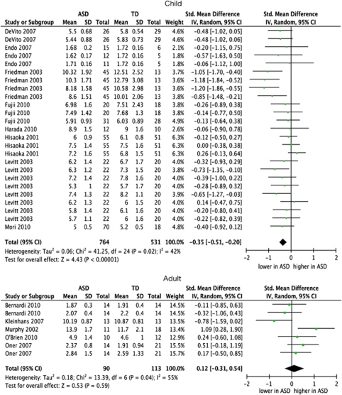
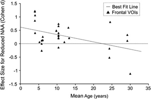
Abnormal trajectory of brain development has been suggested by previous structural magnetic resonance imaging and head circumference findings in autism spectrum disorders (ASDs); however, the neurochemical backgrounds remain unclear. To elucidate neurochemical processes underlying aberrant brain growth in ASD, we conducted a comprehensive literature search and a meta-analysis of (1)H-magnetic resonance spectroscopy ((1)H-MRS) studies in ASD. From the 22 articles identified as satisfying the criteria, means and s.d. of measure of N-acetylaspartate (NAA), creatine, choline-containing compounds, myo-Inositol and glutamate+glutamine in frontal, temporal, parietal, amygdala-hippocampus complex, thalamus and cerebellum were extracted. Random effect model analyses showed significantly lower NAA levels in all the examined brain regions but cerebellum in ASD children compared with typically developed children (n=1295 at the maximum in frontal, P<0.05 Bonferroni-corrected), although there was no significant difference in metabolite levels in adulthood. Meta-regression analysis further revealed that the effect size of lower frontal NAA levels linearly declined with older mean age in ASD (n=844, P<0.05 Bonferroni-corrected). The significance of all frontal NAA findings was preserved after considering between-study heterogeneities (P<0.05 Bonferroni-corrected). This first meta-analysis of (1)H-MRS studies in ASD demonstrated robust developmental changes in the degree of abnormality in NAA levels, especially in frontal lobes of ASD. Previously reported larger-than-normal brain size in ASD children and the coincident lower-than-normal NAA levels suggest that early transient brain expansion in ASD is mainly caused by an increase in non-neuron tissues, such as glial cell proliferation.
Figures
References
-
- Frith U, Happe F, Siddons F. Autism and theory of mind in everyday life. Soc Dev. 1994;3:108–124. - PubMed
-
- Frith U, Happe F. Language and communication in autistic disorders. Phil Trans R Soc B Biol Sci. 1994;346:97–104. - PubMed
-
- Redcay E, Courchesne E. When is the brain enlarged in autism? A meta-analysis of all brain size reports. Biol Psychiatry. 2005;58:1–9. - PubMed
-
- Courchesne E, Carper R, Akshoomoff N. Evidence of brain overgrowth in the first year of life in autism. JAMA. 2003;290:337–344. - PubMed
Publication types
MeSH terms
Substances
LinkOut - more resources
Full Text Sources
