. 2012 Sep;21(9):1748-57.
doi: 10.1007/s00586-012-2442-5. Epub 2012 Jul 26.

Heme oxygenase-1 modulates degeneration of the intervertebral disc after puncture in Bach 1 deficient mice

Affiliations

Heme oxygenase-1 modulates degeneration of the intervertebral disc after puncture in Bach 1 deficient mice

Ryo Ohta et al. Eur Spine J. 2012 Sep.

Abstract

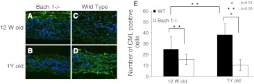
Purpose: Intervertebral disc degeneration is considered to be a major feature of low back pain. Furthermore, oxidative stress has been shown to be an important factor in degenerative diseases such as osteoarthritis and is considered a cause of intervertebral disc degeneration. The purpose of this study was to clarify the correlation between oxidative stress and intervertebral disc degeneration using Broad complex-Tramtrack-Bric-a-brac and cap'n'collar homology 1 deficient (Bach 1-/-) mice which highly express heme oxygenase-1 (HO-1). HO-1 protects cells from oxidative stress.

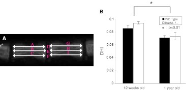
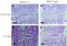
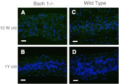
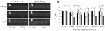
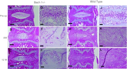
Methods: Caudal discs of 12-week-old and 1-year-old mice were evaluated as age-related models. Each group and period, 5 mice (a total of 20 mice, a total of 20 discs) were evaluated as age-related model. C9-C10 caudal discs in 12-week-old Bach 1-/- and wild-type mice were punctured using a 29-gauge needle as annulus puncture model. Each group and period, 5 mice (a total of 60 mice, a total of 60 discs) were evaluated. The progress of disc degeneration was evaluated at pre-puncture, 1, 2, 4, 8 and 12 weeks post-puncture. Radiographic, histologic and immunohistologic analysis were performed to compare between Bach 1-/- and wild-type mice.

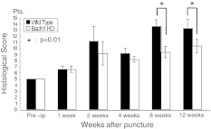
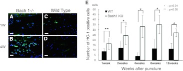
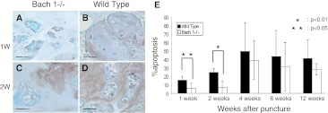
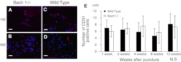
Results: In the age-related model, there were no significant differences between Bach 1-/- and wild-type mice radiologically and histologically. However, in the annulus puncture model, histological scoring revealed significant difference at 8 and 12 weeks post-puncture. The number of HO-1 positive cells was significantly greater in Bach 1-/- mice at every period. The apoptosis rate was significantly lower at 1 and 2 weeks post-puncture in Bach 1-/- mice.

Conclusions: Oxidative stress prevention may avoid the degenerative process of the intervertebral disc after puncture, reducing the number of apoptosis cells. High HO-1 expression may also inhibit oxidative stress and delay the process of intervertebral disc degeneration.


Figures

Fig. 1
Fig. 1
On lateral radiographs, the vertebral body height and intervertebral disc height were measured. Disc height index (DHI) was calculated by averaging the height of the anterior, middle, and posterior portions of the intervertebral disc and dividing that by the average of adjacent vertebral body heights (a). DHI = 2 × (D + E + F)/(A + B + C + G + H + I). Change of DHI of age-related model (b). At the age of 1 year, the DHI significantly decreased compared with that at 12 weeks. No significant difference was observed between Bach 1−/− mice and wild-type mice in both periods
Fig. 2
Fig. 2
Histological changes of the age-related model. Overview of histological changes in the intervertebral discs of 12-week-old mice (a, c). The notochordal cells were uniformly spread in the nucleus pulposus, and the notochordal cells gathered at the center of the nucleus pulposus at 1 year (b, d). Scale bar = 200 μm (ad)
Fig. 3
Fig. 3
Distribution of CML in the center of the intervertebral disc. Typical examples showing immunofluorescence of CML (green) and nucleus (blue) in sagittal sections of intervertebral discs from Bach 1−/− (a, b) and wild-type (c, d) mice. Scale bar = 40 μm. The number of CML positive cells in the center of the intervertebral disc in one visual field of ×400 (e). The CML-positive cells increased in wild-type mice, but in Bach 1−/− mice, CML positive cells were less than that in 12-week- and 1-year-old wild-type mice. CML-positive cells significantly increased in wild-type mice, but in Bach 1−/− mice, the CML-positive cells were small in number
Fig. 4
Fig. 4
Distribution of HO-1 protein in the center of the intervertebral disc. Typical examples showing immunofluorescence of HO-1 (green) and nucleus (blue) in sagittal sections of intervertebral discs from Bach 1−/− (a, b) and wild-type (c, d) mice. Scale bar = 40 μm
Fig. 5
Fig. 5
Lateral radiographs of annulus needle puncture model. The disc height of punctured intervertebral disc (C9/10) decreased at 1 week (b, e) to 12 weeks (c, f) compared with pre-operation (a, d) and control (C8/9). Change of % disc height index (%DHI) of the annulus needle puncture model (g). A significant decrease of %DHI was observed compared with the %DHI of the control disc (C8–C9). In the control intervertebral discs and punctured intervertebral disc, no significant difference was observed between Bach 1−/− mice and wild-type mice at every period
Fig. 6
Fig. 6
Histological changes of the annulus needle puncture model. Overview of histological changes in the intervertebral discs by pre-operative Hematoxylin and eosin staining, 8 and 12 weeks after puncture in Bach 1−/− mice (af) and wild-type mice (gl). The histological changes are clear in both groups, and less degenerative change can be observed in Bach 1−/− mice at 8 and 12 weeks after puncture. Scale bar = 200 μm (ac, gi) and Scale bar = 40 μm (df, jl)
Fig. 7
Fig. 7
Changes in the histological score of the punctured disc. The histological score increased from pre-operation to 12 weeks after puncture. The histological score of Bach 1−/− mice at 8 and 12 weeks after puncture was significantly lower than that of wild-type mice (p < 0.01)
Fig. 8
Fig. 8
Distribution of HO-1 protein in the center of the intervertebral disc. Typical examples showing immunofluorescence of HO-1 (green) and nucleus (blue) in sagittal sections of intervertebral discs from Bach 1−/− (a, b) and wild-type (c, d) mice. Scale bar = 40 μm. The number of HO-1 positive cells in the center of the intervertebral disc in one visual field of ×400 (e). From 1 week to 12 weeks, the number of HO-1 positive cells in Bach 1−/− mice was significantly greater than that of wild-type mice at any period of time
Fig. 9
Fig. 9
Detection of apoptosis cells. TUNEL-stained sections at 1 and 2 weeks after puncture (ad). Apoptosis cells were stained brown and non-apoptosis cells were stained blue. The number of apoptosis cells increased after puncture in wild-type mice more than in Bach 1−/− mice. Scale bar = 40 μm. The apoptosis rate of Bach 1−/− mice was significantly lower than that of wild-type mice at 1 week (p < 0.01) and 2 weeks (p < 0.05) (e). No significant difference was observed from 4 to 12 weeks after puncture between Bach 1−/− mice and wild-type mice
Fig. 10
Fig. 10
Distribution of CD31 in the center of the intervertebral disc at 1 and 4 weeks after puncture. Typical examples showing immunofluorescence of CD31 (red) and nucleus (blue) in sagittal sections of intervertebral discs from Bach 1−/− (a, b) and wild-type (c, d) mice. Scale bar = 40 μm. The number of CD31 positive cells in the center of the intervertebral disc in one visual field of ×400 (e). From 1 to 12 weeks, the number of CD31-positive cells in Bach 1−/− mice did not change significantly, and no significant difference was observed between Bach 1−/− mice and wild-type mice

Similar articles

Cited by

References

    1. Kirkaldy-Willis WH, Farfan HF. Instability of the lumbar spine. Clin Orthop Relat Res. 1982;165:110–123. - PubMed
    1. Urban JP, Roberts S. Degeneration of the intervertebral disc. Arthritis Res Ther. 2003;5:120–130. doi: 10.1186/ar629. - DOI - PMC - PubMed
    1. Tanaka N, An HS, Lim TH, et al. The relationship between disc degeneration and flexibility of the lumbar spine. Spine J. 2001;1:47–56. doi: 10.1016/S1529-9430(01)00006-7. - DOI - PubMed
    1. An HS, Masuda K, Inoue N. Intervertebral disc degeneration: biological and biomechanical factors. J Orthop Sci. 2006;11:541–552. doi: 10.1007/s00776-006-1055-4. - DOI - PMC - PubMed
    1. Seki S, Kawaguchi Y, Chiba K, et al. A functional SNP in CILP, encoding cartilage intermediate layer protein, is associated with susceptibility to lumbar disc disease. Nat Genet. 2005;37:607–612. doi: 10.1038/ng1557. - DOI - PubMed

Publication types

Substances