

UMMPerfusion: an open source software tool towards quantitative MRI perfusion analysis in clinical routine
- PMID: 22832894
- PMCID: PMC3597952
- DOI: 10.1007/s10278-012-9510-6
UMMPerfusion: an open source software tool towards quantitative MRI perfusion analysis in clinical routine
Abstract
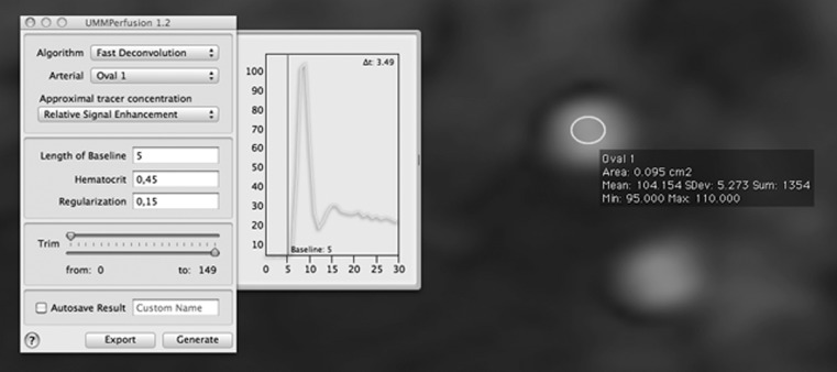
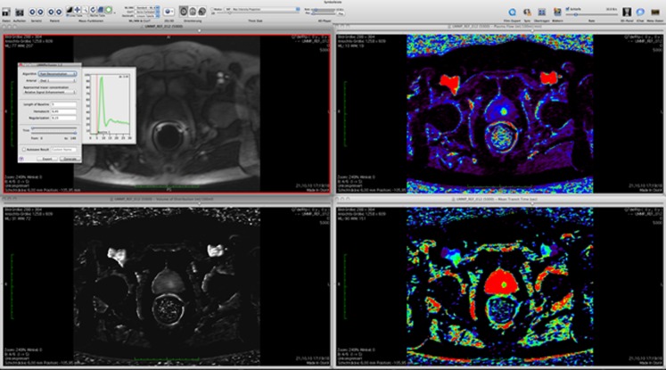
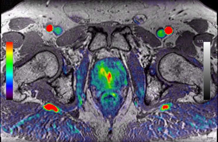
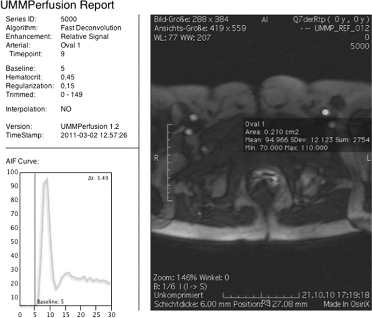
To develop a generic Open Source MRI perfusion analysis tool for quantitative parameter mapping to be used in a clinical workflow and methods for quality management of perfusion data. We implemented a classic, pixel-by-pixel deconvolution approach to quantify T1-weighted contrast-enhanced dynamic MR imaging (DCE-MRI) perfusion data as an OsiriX plug-in. It features parallel computing capabilities and an automated reporting scheme for quality management. Furthermore, by our implementation design, it could be easily extendable to other perfusion algorithms. Obtained results are saved as DICOM objects and directly added to the patient study. The plug-in was evaluated on ten MR perfusion data sets of the prostate and a calibration data set by comparing obtained parametric maps (plasma flow, volume of distribution, and mean transit time) to a widely used reference implementation in IDL. For all data, parametric maps could be calculated and the plug-in worked correctly and stable. On average, a deviation of 0.032 ± 0.02 ml/100 ml/min for the plasma flow, 0.004 ± 0.0007 ml/100 ml for the volume of distribution, and 0.037 ± 0.03 s for the mean transit time between our implementation and a reference implementation was observed. By using computer hardware with eight CPU cores, calculation time could be reduced by a factor of 2.5. We developed successfully an Open Source OsiriX plug-in for T1-DCE-MRI perfusion analysis in a routine quality managed clinical environment. Using model-free deconvolution, it allows for perfusion analysis in various clinical applications. By our plug-in, information about measured physiological processes can be obtained and transferred into clinical practice.
Figures
References
-
- Scherr MK, Seitz M, Muller-Lisse UG, Ingrisch M, Reiser MF, Muller-Lisse UL. MR-perfusion (MRP) and diffusion-weighted imaging (DWI) in prostate cancer: quantitative and model-based gadobenate dimeglumine MRP parameters in detection of prostate cancer. Eur J Radiol. 2010;76:359–366. doi: 10.1016/j.ejrad.2010.04.023. - DOI - PubMed
Publication types
MeSH terms
LinkOut - more resources
Full Text Sources
