Genetic inhibition of fibroblast growth factor receptor 1 in knee cartilage attenuates the degeneration of articular cartilage in adult mice
- PMID: 22833219
- PMCID: PMC3690192
- DOI: 10.1002/art.34645
Genetic inhibition of fibroblast growth factor receptor 1 in knee cartilage attenuates the degeneration of articular cartilage in adult mice
Abstract
Objective: Fibroblast growth factor (FGF) family members are involved in the regulation of articular cartilage homeostasis. The aim of this study was to investigate the function of FGF receptor 1 (FGFR-1) in the development of osteoarthritis (OA) and its underlying mechanisms.
Methods: FGFR-1 was deleted from the articular chondrocytes of adult mice in a cartilage-specific and tamoxifen-inducible manner. Two OA models (aging-associated spontaneous OA, and destabilization-induced OA), as well as an antigen-induced arthritis (AIA) model, were established and tested in Fgfr1-deficient and wild-type (WT) mice. Alterations in cartilage structure and the loss of proteoglycan were assessed in the knee joints of mice of either genotype, using these 3 arthritis models. Primary chondrocytes were isolated and the expression of key regulatory molecules was assessed quantitatively. In addition, the effect of an FGFR-1 inhibitor on human articular chondrocytes was examined.
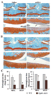
Results: The gross morphologic features of Fgfr1-deficient mice were comparable with those of WT mice at both the postnatal and adult stages. The articular cartilage of 12-month-old Fgfr1-deficient mice displayed greater aggrecan staining compared to 12-month-old WT mice. Fgfr1 deficiency conferred resistance to the proteoglycan loss induced by AIA and attenuated the development of cartilage destruction after surgically induced destabilization of the knee joint. The chondroprotective effect of FGFR-1 inhibition was largely associated with decreased expression of matrix metalloproteinase 13 (MMP-13) and up-regulation of FGFR-3 in mouse and human articular chondrocytes.
Conclusion: Disruption of FGFR-1 in adult mouse articular chondrocytes inhibits the progression of cartilage degeneration. Down-regulation of MMP-13 expression and up-regulation of FGFR-3 levels may contribute to the phenotypic changes observed in Fgfr1-deficient mice.
Copyright © 2012 by the American College of Rheumatology.
Figures


Comment in
-
Explaining the fibroblast growth factor paradox in osteoarthritis: lessons from conditional knockout mice.Arthritis Rheum. 2012 Dec;64(12):3835-8. doi: 10.1002/art.34648. Arthritis Rheum. 2012. PMID: 22833278 No abstract available.
Similar articles
-
Fibroblast Growth Factor Receptor 3 Inhibits Osteoarthritis Progression in the Knee Joints of Adult Mice.Arthritis Rheumatol. 2016 Oct;68(10):2432-43. doi: 10.1002/art.39739. Arthritis Rheumatol. 2016. PMID: 27159076
-
Fibroblast growth factor receptor 1 is principally responsible for fibroblast growth factor 2-induced catabolic activities in human articular chondrocytes.Arthritis Res Ther. 2011 Aug 11;13(4):R130. doi: 10.1186/ar3441. Arthritis Res Ther. 2011. Retraction in: Arthritis Res Ther. 2024 Aug 7;26(1):149. doi: 10.1186/s13075-024-03381-y. PMID: 21835001 Free PMC article. Retracted.
-
A novel FGFR1-binding peptide attenuates the degeneration of articular cartilage in adult mice.Osteoarthritis Cartilage. 2018 Dec;26(12):1733-1743. doi: 10.1016/j.joca.2018.08.012. Epub 2018 Sep 8. Osteoarthritis Cartilage. 2018. PMID: 30201491
-
Fibroblast growth factor control of cartilage homeostasis.J Cell Biochem. 2013 Apr;114(4):735-42. doi: 10.1002/jcb.24418. J Cell Biochem. 2013. PMID: 23060229 Free PMC article. Review.
-
Regulatory Mechanisms of Prg4 and Gdf5 Expression in Articular Cartilage and Functions in Osteoarthritis.Int J Mol Sci. 2022 Apr 23;23(9):4672. doi: 10.3390/ijms23094672. Int J Mol Sci. 2022. PMID: 35563063 Free PMC article. Review.
Cited by
-
Regulation of FGF-2, FGF-18 and Transcription Factor Activity by Perlecan in the Maturational Development of Transitional Rudiment and Growth Plate Cartilages and in the Maintenance of Permanent Cartilage Homeostasis.Int J Mol Sci. 2022 Feb 9;23(4):1934. doi: 10.3390/ijms23041934. Int J Mol Sci. 2022. PMID: 35216048 Free PMC article. Review.
-
Targeting local lymphatics to ameliorate heterotopic ossification via FGFR3-BMPR1a pathway.Nat Commun. 2021 Jul 19;12(1):4391. doi: 10.1038/s41467-021-24643-2. Nat Commun. 2021. PMID: 34282140 Free PMC article.
-
Aging processes and the development of osteoarthritis.Curr Opin Rheumatol. 2013 Jan;25(1):108-13. doi: 10.1097/BOR.0b013e32835a9428. Curr Opin Rheumatol. 2013. PMID: 23080227 Free PMC article. Review.
-
Fibroblast growth factor signalling in osteoarthritis and cartilage repair.Nat Rev Rheumatol. 2020 Oct;16(10):547-564. doi: 10.1038/s41584-020-0469-2. Epub 2020 Aug 17. Nat Rev Rheumatol. 2020. PMID: 32807927 Review.
-
Chaperonin 60 regulation of SOX9 ubiquitination mitigates the development of knee osteoarthritis.J Mol Med (Berl). 2016 Jul;94(7):755-69. doi: 10.1007/s00109-016-1422-3. Epub 2016 Apr 27. J Mol Med (Berl). 2016. PMID: 27118120
References
Publication types
MeSH terms
Substances
Grants and funding
LinkOut - more resources
Full Text Sources
Molecular Biology Databases
Miscellaneous

