

Knockdown of IKK1/2 promotes differentiation of mouse embryonic stem cells into neuroectoderm at the expense of mesoderm
- PMID: 22833419
- PMCID: PMC3505544
- DOI: 10.1007/s12015-012-9402-7
Knockdown of IKK1/2 promotes differentiation of mouse embryonic stem cells into neuroectoderm at the expense of mesoderm
Abstract
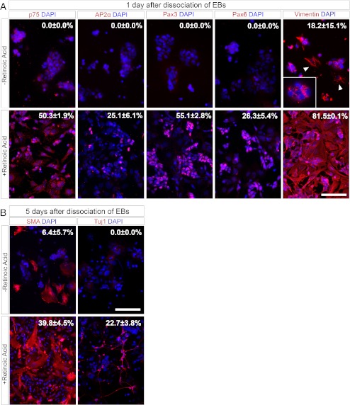
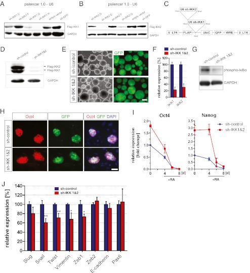
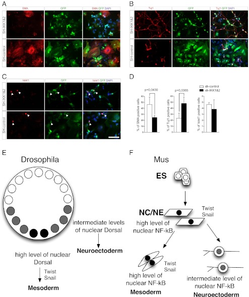
Activation of nuclear factor kappa B (NF-κB) is accomplished by a specific kinase complex (IKK-complex), phosphorylating inhibitors of NF-κB (IκB). In embryonic stem cells (ESCs), NF-κB signaling causes loss of pluripotency and promotes differentiation towards a mesodermal phenotype. Here we show that NF-κB signaling is involved in cell fate determination during retinoic acid (RA) mediated differentiation of ESCs. Knockdown of IKK1 and IKK2 promotes differentiation of ESCs into neuroectoderm at the expense of neural crest derived myofibroblasts. Our data indicate that RA is not only able to induce neuronal differentiation in vitro but also drives ESCs into a neural crest cell lineage represented by differentiation towards peripheral neurons and myofibroblasts. The NC is a transiently existing, highly multipotent embryonic cell population generating a wide range of different cell types. During embryonic development the NC gives rise to distinct precursor lineages along the anterior-posterior axis determining differentiation towards specific derivates. Retinoic acid (RA) signaling provides essential instructive cues for patterning the neuroectoderm along the anterior-posterior axis. The demonstration of RA as a sufficient instructive signal for the differentiation of pluripotent cells towards NC and the involvement of NF-κB during this process provides useful information for the generation of specific NC-lineages, which are valuable for studying NC development or disease modeling.
Figures

Similar articles
-
Inhibition of IKK/NF-κB Signaling Enhances Differentiation of Mesenchymal Stromal Cells from Human Embryonic Stem Cells.Stem Cell Reports. 2016 Apr 12;6(4):456-465. doi: 10.1016/j.stemcr.2016.02.006. Epub 2016 Mar 10. Stem Cell Reports. 2016. PMID: 26972683 Free PMC article.
-
Heparan sulfate facilitates FGF and BMP signaling to drive mesoderm differentiation of mouse embryonic stem cells.J Biol Chem. 2012 Jun 29;287(27):22691-700. doi: 10.1074/jbc.M112.368241. Epub 2012 May 3. J Biol Chem. 2012. PMID: 22556407 Free PMC article.
-
IκB kinase/nuclear factor κB-dependent insulin-like growth factor 2 (Igf2) expression regulates synapse formation and spine maturation via Igf2 receptor signaling.J Neurosci. 2012 Apr 18;32(16):5688-703. doi: 10.1523/JNEUROSCI.0111-12.2012. J Neurosci. 2012. PMID: 22514330 Free PMC article.
-
The issue of the multipotency of the neural crest cells.Dev Biol. 2018 Dec 1;444 Suppl 1:S47-S59. doi: 10.1016/j.ydbio.2018.03.024. Epub 2018 Mar 31. Dev Biol. 2018. PMID: 29614271 Review.
-
The stem cells of the neural crest.Cell Cycle. 2008 Apr 15;7(8):1013-9. doi: 10.4161/cc.7.8.5641. Epub 2008 Jan 24. Cell Cycle. 2008. PMID: 18414040 Review.
Cited by
-
Identifying the potential transcriptional regulatory network in Hirschsprung disease by integrated analysis of microarray datasets.World J Pediatr Surg. 2023 Apr 17;6(2):e000547. doi: 10.1136/wjps-2022-000547. eCollection 2023. World J Pediatr Surg. 2023. PMID: 37082700 Free PMC article.
-
Cervical cancer stem cells and other leading factors associated with cervical cancer development.Oncol Lett. 2019 Oct;18(4):3423-3432. doi: 10.3892/ol.2019.10718. Epub 2019 Aug 6. Oncol Lett. 2019. PMID: 31516560 Free PMC article. Review.
-
Simple method for sub-diffraction resolution imaging of cellular structures on standard confocal microscopes by three-photon absorption of quantum dots.PLoS One. 2013 May 21;8(5):e64023. doi: 10.1371/journal.pone.0064023. Print 2013. PLoS One. 2013. PMID: 23700448 Free PMC article.
-
Genome-wide gene by lead exposure interaction analysis identifies UNC5D as a candidate gene for neurodevelopment.Environ Health. 2017 Jul 28;16(1):81. doi: 10.1186/s12940-017-0288-3. Environ Health. 2017. PMID: 28754176 Free PMC article.
-
The Transcription Factor NF-κB in Stem Cells and Development.Cells. 2021 Aug 10;10(8):2042. doi: 10.3390/cells10082042. Cells. 2021. PMID: 34440811 Free PMC article. Review.
References
-
- Lüningschrör, P., Stöcker, B., Kaltschmidt, B., Kaltschmidt, C. (2012). miR-290 cluster modulates pluripotency by repressing canonical NF-kappaB signaling. Stem Cells. - PubMed
Publication types
MeSH terms
Substances
LinkOut - more resources
Full Text Sources
