. 2012 Nov 1;21(21):4602-14.
doi: 10.1093/hmg/dds299. Epub 2012 Jul 23.

Entorhinal cortical neurons are the primary targets of FUS mislocalization and ubiquitin aggregation in FUS transgenic rats

Affiliations

Entorhinal cortical neurons are the primary targets of FUS mislocalization and ubiquitin aggregation in FUS transgenic rats

Cao Huang et al. Hum Mol Genet. .

Abstract

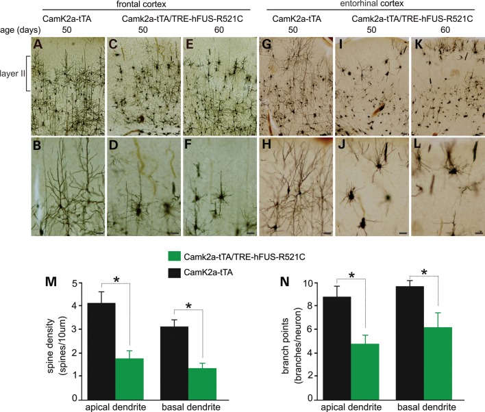
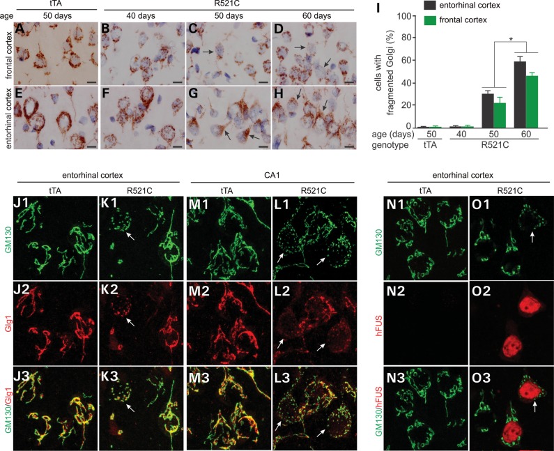
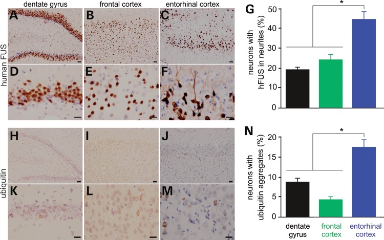
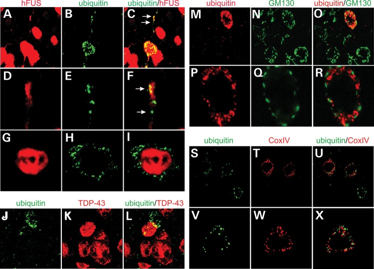
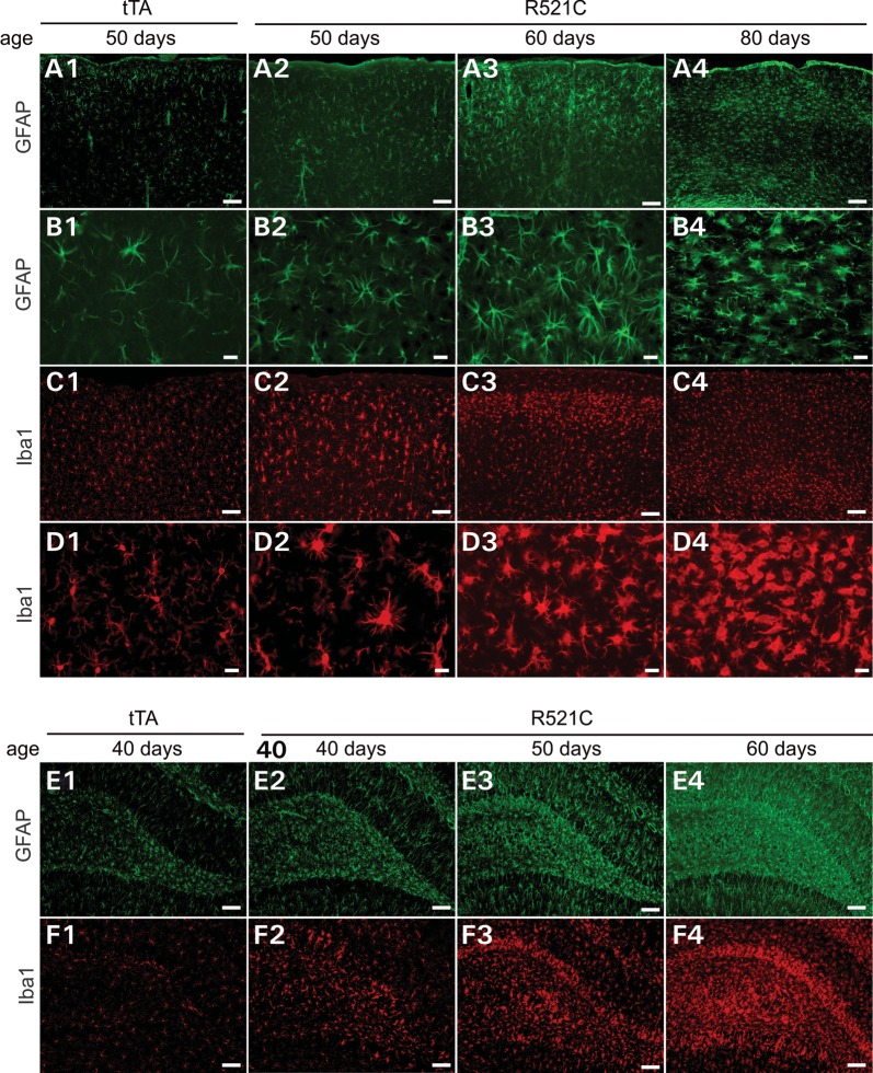
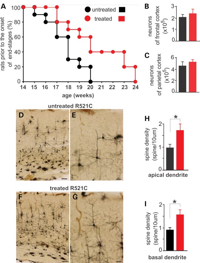
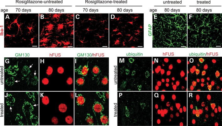
Ubiquitin-positive inclusion containing Fused in Sarcoma (FUS) defines a new subtype of frontotemporal lobar degeneration (FTLD). FTLD is characterized by progressive alteration in cognitions and it preferentially affects the superficial layers of frontotemporal cortex. Mutation of FUS is linked to amyotrophic lateral sclerosis and to motor neuron disease with FTLD. To examine FUS pathology in FTLD, we developed the first mammalian animal model expressing human FUS with pathogenic mutation and developing progressive loss of memory. In FUS transgenic rats, ubiquitin aggregation and FUS mislocalization were developed primarily in the entorhinal cortex of temporal lobe, particularly in the superficial layers of affected cortex. Overexpression of mutant FUS led to Golgi fragmentation and mitochondrion aggregation. Intriguingly, aggregated ubiquitin was not colocalized with either fragmented Golgi or aggregated mitochondria, and neurons with ubiquitin aggregates were deprived of endogenous TDP-43. Agonists of peroxisome proliferator-activated receptor gamma (PPAR-γ) possess anti-glial inflammation effects and are also shown to preserve the dendrite and dendritic spines of cortical neurons in culture. Here we show that rosiglitazone, a PPAR-γ agonist, rescued the dendrites and dendritic spines of neurons from FUS toxicity and preserved rats' spatial memory. Our FUS transgenic rats would be useful to the mechanistic study of cortical dementia in FTLD. As rosiglitazone is clinically used to treat diabetes, our results would encourage immediate application of PPAR-γ agonists in treating patients with cortical dementia.


Figures

Figure 1.
Figure 1.
Restricted expression of the reporter gene LacZ in the forebrain of transgenic rats. (A) The schematic shows a tetracycline-regulated gene expression system. The promoter of mouse Camk2a directs the synthesis of tTA, which is subject to the regulation by Dox. Free tTA binds to tetracycline-responsive elements (TRE) and activate the LacZ transgene. (B) A reconstructed photo shows the profile of LacZ expression on the sagittal section of a rat brain. Tissue sections were taken from a Camk2a-tTA/TRE-LacZ double-transgenic 4-week-old rat. Individual photos were assembled with Photoshop to demonstrate the profile of X-gal staining. (C and D) X-gal staining shows the expression patterns of the LacZ transgene in the cortex (C) and CA1 region (D). Tissue sections were first stained with X-gal and were further stained with an antibody to myelin basic protein. (E) A cross-section of cervical spinal cord shows that corticospinal tracks were stained with X-gal. Scale bars: (B) 1 mm; (C and D) 100 µm; (E) 200 µm.
Figure 2.
Figure 2.
Camk2a-tTA activates human FUS transgene selectively in the neurons of a rat forebrain. (AO) Double-labeling fluorescence staining reveals that human FUS (hFUS) is colocalized with the neuronal marker NeuN and is not colocalized with the glia markers (GFAP for astrocytes and APC for oligodendrocytes) in the dentate gyrus (A, B and G–I) and cortex (D–F and J–O) of Camk2a-tTA/TRE-FUSR521C double-transgenic rats. Transgenic rats were given Dox during embryonic and postnatal development and were deprived of Dox at 30 days of age. The hippocampus was taken from a 50-day-old rat (20 days off Dox), but the cortex was taken from a 60-day-old rat (30 days off Dox). All photos were taken at identical magnification.
Figure 3.
Figure 3.
Restricted overexpression of mutant FUS in a rat forebrain causes progressive loss of neurons and memory. (A) The diagram shows a strategy to induce the expression of mutant human FUS (hFUS) (R521C) in Camk2a-tTA/TRE-FUSR521C transgenic rats. Breeding rats and their offspring were provided with Dox in drinking water and the transgenic offspring were deprived of Dox by the age of 30 days. After Dox was withdrawn, hFUS began to express from the zero level to the maximal levels (plateau). (BI) Immunofluorescent staining revealed the gradual expression of mutant hFUS in Camk2a-tTA/TRE-FUSR521C double-transgenic rats (R521C), but not in Camk2a-tTA single-transgenic rats (tTA). (J) The Barnes maze revealed the reduction of spatial memory in mutant FUS transgenic rats (R521C), but not in Camk2a-tTA single-transgenic rats (tTA) or in non-transgenic rats (NT). Rat's spatial memory was measured with Banes maze once a week. Data are means + SEM (n = 8–10). (KR) Cresyl violet staining revealed the progressive loss of neurons in Camk2a-tTA/TRE-FUSR521C double-transgenic rats (R521C). Scale bars: (K–N) 100µm; and (O–R) 20µm. (S and T) Stereological cell counting estimated the number of neurons in specified brain regions in the Camk2a-tTA/TRE-FUSR521C double-transgenic rats (R521C) and in the Camk2a-tTA single-transgenic rats (tTA). Data are means SEM (n = 6–7). *P < 0.05.
Figure 4.
Figure 4.
Neurites and spines are lost prior to neuron loss. (AL) Representative photos of Golgi staining showing that affected neurons displayed contracted neurites particularly in the layer II of frontal and entorhinal cortexes in mutant FUS transgenic rats. All scale bars: 40 µm. (M and N) Spine densities and branch points were quantified for five neurons in the frontal cortex of individual rats. Data are means + SEM (n = 4). *P < 0.05.
Figure 5.
Figure 5.
Overexpression of mutant human FUS leads to Golgi fragmentation. (AH) Immunostaining reveals fragmented Golgi apparatuses in the Camk2a-tTA/TRE-FUSR521C double-transgenic rat (R521C), but not in the Camk2a-tTA single-transgenic rat (tTA). Coronal sections (12 µm) of the forebrain were stained with an antibody to GM130. Scale bars: 10 µm. (I) The cells with fragmented Golgi were quantified. Ten images (×100) were examined for Golgi fragmentation in the frontal and entorhinal cortexes of individual rats. Data are means + SEM (n = 5). *P < 0.05. (J–L) Confocal microscopy reveals that the cis (stained with GM130) and trans (stained with Glg1) faces of Golgi were fragmented in affected cells. Arrows point to fragmented Golgi. (N and O) Confocal microscopy reveals that Golgi fragmentation occurred in neurons expressing mutant human FUS (R521). Coronal sections (12 µm) of the forebrain were stained with GM130 and with Glg1 or human FUS antibodies and Z-stacks of confocal images were projected to construct Golgi apparatuses (J–O).
Figure 6.
Figure 6.
FUS mislocalization and ubiquitin aggregation primarily develop in the entorhinal cortex of transgenic rats. (AF) Immunostaining showing that mutant human FUS was mislocalized to the neurites of some neurons in the Camk2a-tTA/TRE-FUSR521C double-transgenic rats. (G) Neurons with human FUS in the neurites were quantified for selected brain regions. Data are means + SEM (n = 5). (HM) Immunostaining showing that ubiquitin formed aggregates in some neurons. (N) Neurons with ubiquitin aggregates were quantified for selected brain regions. Data are means + SEM (n = 4). Neurons were examined for FUS mislocalization (human FUS in the neurites) and ubiquitin aggregation on six images (×40) of each selected brain region for individual rats at the age of 50 days. FUS mislocalization is defined as a neuron with human FUS in a neurite that is longer than the diameter of the cell body. *P < 0.05. Scale bars: (A–C and H–J) 40 µm; (D–F and K–M) 20 µm.
Figure 7.
Figure 7.
Ubiquitin aggregation and Golgi fragmentation co-exist in affected neurons. (AI) Confocal microscopy reveals that aggregated ubiquitin was not physically colocalized with human FUS in the entorhinal cortical neurons of transgenic rats. Z-stacks images were projected to show the profile of ubiquitin aggregation and human FUS mislocalization (A–C). Single-scanned images (thickness: 2 µm) showed the localization of ubiquitin aggregates and human FUS. (JL) Confocal microscopy reveals that neurons with ubiquitin aggregates were depleted of rat endogenous TDP-43. (MR) Confocal microscopy reveals that ubiquitin aggregates coexisted, but were not colocalized, with Golgi fragments in the same affected neurons. Z-stack images were projected to show the profile of ubiquitin aggregation and Golgi fragmentation (M–O). Single-scanned images (2 µm) showed the localization of ubiquitin aggregates and Golgi fragments (P–R). (SX) Confocal microscopy reveals that ubiquitin aggregates were not colocalized with damaged mitochondria immunostained of Cox-IV. Lateral entorhinal cortex was taken from a Camk2a-tTA/TRE-FUSR521C transgenic rat at the age of 60 days.
Figure 8.
Figure 8.
Astrocytes and microglia are overtly activated in mutant FUS transgenic rats. (AF) Immunostaining for GFAP (a marker of astrocyte) and Iba1 (a marker of microglia) revealed that astrocytes and microglia were activated in the cortex (A–D) and dentate gyrus (E and F) of Camk2a-tTA/TRE-FUSR521C double-transgenic rat (R521C) when compared with Camk2a-tTA single-transgenic rat (tTA). The glial cells were first activated in the layers II and III of cortex (A3 and C2–3). Scale bars: (A1–4, C1–4, E1–4 and F1–4): 100 µm; (B1–4 and D1–4): 20 µm.
Figure 9.
Figure 9.
Rosiglitazone treatment preserves neurites and memory in mutant FUS transgenic rats. (A) The graph shows disease progression in Camk2a-tTA/TRE-FUSR521C double-transgenic rats that were treated with (treated) or without (untreated) rosiglitazone. Rats' spatial memory was weekly determined in a Barnes maze and disease stages were determined accordingly. Disease onset was defined as unrecoverable increase in the latency to find an escaping hole in a Barnes maze, and disease end-stages were defined as the escaping latency >60 s. Rats that reached the disease end-stages were counted and terminated. Rats received rosiglitazone treatment from disease onset onwards. Data are means + SEM (n = 15). (B and C) Stereological cell counting reveals no difference in the number of neurons between rosiglitazone-treated and untreated FUS transgenic rats. Data are means + SEM (n = 5). (DG) Golgi staining reveals that neurites were preserved in rosiglitazone-treated rats (F and G) when compared with the untreated rats (D and E). Scale bars: (D and F) 100 µm; (E and G) 40 µm. (H and I) Spine density was quantified for five neurons per rat. Data are means + SEM (n = 4). *P < 0.05.
Figure 10.
Figure 10.
Rosiglitazone mitigates glial reaction and ubiquitin aggregation. (AF) Immunofluorescent staining reveals microglial (Iba-1: A–D) and astroglial (GFAP: E and F) reaction in rosiglitazone-treated (C, D and F) and -untreated (A, B and E) rats. Scale bars: 20 µm. (GR) Confocal microscopy reveals that rosiglitazone moderately mitigated Golgi fragmentation (G–L) and ubiquitin aggregation (M–R) in rosiglitazone-treated rats (J–L and P–R) compared with untreated rats (G–I and M–O). Camk2a-tTA/TRE-FUSR521C double-transgenic rats received rosiglitazone treatment from disease onset onwards and were terminated at the age of 70 days (A and C) or 80 days (B, D and E–R). Rat entorhinal cortices were analyzed for Golgi fragmentation (GM130) and ubiquitin aggregation. Neurons expressing mutant FUS were identified by immunostaining for human FUS (hFUS). Arrows point to fragmented Golgi apparatus (G and J).

Similar articles

Cited by

References

    1. Galimberti D., Scarpini E. Genetics and biology of Alzheimer's disease and frontotemporal lobar degeneration. Int. J. Clin. Exp. Med. 2010;3:129–143. - PMC - PubMed
    1. Hu W.T., Trojanowski J.Q., Shaw L.M. Biomarkers in frontotemporal lobar degenerations—progress and challenges. Prog. Neurobiol. 2011;95:636–648. - PMC - PubMed
    1. Mackenzie I.R., Foti D., Woulfe J., Hurwitz T.A. Atypical frontotemporal lobar degeneration with ubiquitin-positive, TDP-43-negative neuronal inclusions. Brain. 2008;131:1282–1293. - PubMed
    1. Mackenzie I.R. The neuropathology and clinical phenotype of FTD with progranulin mutations. Acta Neuropathol. 2007;114:49–54. - PubMed
    1. Neumann M., Sampathu D.M., Kwong L.K., Truax A.C., Micsenyi M.C., Chou T.T., Bruce J., Schuck T., Grossman M., Clark C.M., et al. Ubiquitinated TDP-43 in frontotemporal lobar degeneration and amyotrophic lateral sclerosis. Science. 2006;314:130–133. - PubMed

Publication types

MeSH terms