. 2012 Sep;23(18):3731-42.
doi: 10.1091/mbc.E12-03-0172. Epub 2012 Jul 25.

ECM stiffness primes the TGFβ pathway to promote chondrocyte differentiation

Affiliations

ECM stiffness primes the TGFβ pathway to promote chondrocyte differentiation

Jessica L Allen et al. Mol Biol Cell. 2012 Sep.

Abstract

Cells encounter physical cues such as extracellular matrix (ECM) stiffness in a microenvironment replete with biochemical cues. However, the mechanisms by which cells integrate physical and biochemical cues to guide cellular decision making are not well defined. Here we investigate mechanisms by which chondrocytes generate an integrated response to ECM stiffness and transforming growth factor β (TGFβ), a potent agonist of chondrocyte differentiation. Primary murine chondrocytes and ATDC5 cells grown on 0.5-MPa substrates deposit more proteoglycan and express more Sox9, Col2α1, and aggrecan mRNA relative to cells exposed to substrates of any other stiffness. The chondroinductive effect of this discrete stiffness, which falls within the range reported for articular cartilage, requires the stiffness-sensitive induction of TGFβ1. Smad3 phosphorylation, nuclear localization, and transcriptional activity are specifically increased in cells grown on 0.5-MPa substrates. ECM stiffness also primes cells for a synergistic response, such that the combination of ECM stiffness and exogenous TGFβ induces chondrocyte gene expression more robustly than either cue alone through a p38 mitogen-activated protein kinase-dependent mechanism. In this way, the ECM stiffness primes the TGFβ pathway to efficiently promote chondrocyte differentiation. This work reveals novel mechanisms by which cells integrate physical and biochemical cues to exert a coordinated response to their unique cellular microenvironment.


Figures

FIGURE 1:
FIGURE 1:
Chondrocyte differentiation is stiffness sensitive. Primary murine chondrocytes (A) and ATDC5 cells (B) cultured in differentiation media on plastic or polyacrylamide gels of the indicated stiffness demonstrate the greatest increase in Sox9, Col2α1, and aggrecan gene expression on 0.5-MPa substrates, similar to the stiffness of articular cartilage. (C) Alcian blue staining of proteoglycan production, a functional measure of chondrocyte differentiation, reveals a significant 3.1-fold increase in stained area for ATDC5 cells grown on 0.5-MPa substrates relative to cells grown on plastic. Right, high-magnification images of regions outlined on the left. Staining localizes to regions between cells (a single cell perimeter is marked by a dotted line). The small box to the bottom left of each image shows background Alcian blue staining of control cell-free substrates. *p < 0.05.
FIGURE 2:
FIGURE 2:
Substrate stiffness and TGFβ synergize to induce chondrogenic differentiation. Sox9 (A), Col2α1 (B), and aggrecan (C) mRNA expression is greatly induced by TGFβ (5 ng/ml) in ATDC5 cells differentiated for 7 d on 0.5-MPa substrates relative to cells grown on plastic or in the absence of TGFβ. p < 0.01.
FIGURE 3:
FIGURE 3:
Mechanosensitive chondroinduction is mediated by ROCK signaling. Stress fibers, visualized by rhodamine phalloidin (green), are visible in primary chondrocytes (A) and ATDC5 cells (B) with greater frequency and intensity on substrates with stiffnesses of 1.1 MPa or greater. Nuclei are stained with DAPI (red). (C) ROCK inhibition with Y27632 (Y, 10 μM) induces Col2α1 in primary chondrocytes and ATDC5 cells differentiated for 24 h on plastic but represses induction in cells differentiated on 0.5-MPa substrates. Similar results are seen for Sox9 and aggrecan expression. *p < 0.05.
FIGURE 4:
FIGURE 4:
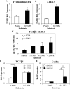
Mechanosensitive, ROCK-dependent induction of TGFβ1 is required for chondroinduction. Within 48 h, TGFβ1 mRNA is induced in primary chondrocytes (A) and ATDC5 cells (B) in a stiffness-dependent manner. (C) TGFβ1 protein produced by ATDC5 cells, as assessed in media by an ELISA, increases with decreasing substrate stiffness. (D) The expression of TGFβ1 mRNA in ATDC5 cells is increased by ROCK inhibition with Y27632 (10 μM) on plastic but is unaffected on 0.5-MPa substrates. (E) Induction of Col2α1 mRNA in ATDC5 cells cultured on a 0.5-MPa gel is impaired by 24-h exposure to SB431542 (5 μM), a chemical antagonist of the TGFβ type I receptor. *p < 0.05, p < 0.01.
FIGURE 5:
FIGURE 5:
Smad3 phosphorylation, localization, and transcriptional activity are sensitive to ECM stiffness. (A) Western analysis of whole-cell lysates from ATDC5 cells cultured as indicated for 24 h reveals that C-terminal Smad3 phosphorylation (top) and total protein (middle) are increased specifically on a 0.5-MPa substrate, whereas β-actin levels (bottom) remain unchanged. (B) Immunofluorescence microscopy allows visualization of stiffness and TGFβ-sensitive Smad2/3 localization (green) relative to DAPI-stained nuclei (red). (C) Relative to cells grown on plastic, a greater percentage of primary chondrocytes and ATDC5 cells have nuclear Smad2/3 when grown on 0.5-MPa substrates, particularly in the absence added TGFβ. (D) In transiently transfected ATDC5 cells grown for 48 h on 0.5-MPa substrates, the activity of the TGFβ-responsive SBE-luciferase promoter reporter construct is increased relative to cells grown on plastic. Luciferase activity is normalized to expression of β-galactosidase from a cotransfected control construct. *p < 0.05, p < 0.01.
FIGURE 6:
FIGURE 6:
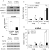
Synergy between TGFβ and ECM stiffness requires p38 MAPK but not Smad3. (A) Western analysis reveals no substrate stiffness-dependent effect on TGFβ-inducible Smad3 phosphorylation. (B) Smad3 shRNA treatment achieves 70% reduction of Smad3 mRNA relative to ATDC5 cells expressing a scrambled shRNA. (C) shSmad3 impairs the stiffness-sensitive induction of Col2α1 mRNA but not the synergistic induction of Col2α1 by TGFβ (5 ng/ml, 24 h) and a 0.5-MPa substrate. (D) Western analysis of phospho-p38 and p38 total protein reveals increased p38 phosphorylation in ATDCs cultured for 24 h on a 0.5-MPa substrate or in the presence of TGFβ (5 ng/ml, 45 min). (E) Synergistic induction of Col2α1 mRNA by a 0.5-MPa substrate and TGFβ is greatly reduced upon inhibition of p38 activity by SB203580 (10 μM) before a 1-h TGFβ treatment. p < 0.01.
FIGURE 7:
FIGURE 7:
Schematic of the mechanism of chondrocyte integration of cues provided by ECM stiffness and TGFβ. 1) Chondrocyte differentiation is specifically induced on 0.5-MPa substrates. 2) This response is mediated through the ROCK pathway. 3) Autocrine TGFβ1 expression is required for chondroinduction, the expression of which is inhibited on stiffer substrates through a ROCK-dependent mechanism. 4) Whereas Smad3 phosphorylation and nuclear localization are increased on 0.5-MPa substrates, softer substrates that express autocrine TGFβ fail to activate this downstream effector. 5) Exogenously added TGFβ elicits a synergistic response in combination with 0.5-MPa substrates. 6) This synergistic interaction acts through a p38-MAPK–dependent mechanism, possibly through TGFβ activation of TAK1.

References

    1. Aigner T, Kurz B, Fukui N, Sandell L. Roles of chondrocytes in the pathogenesis of osteoarthritis. Curr Opin Rheumatol. 2002;14:578–584. - PubMed
    1. Akizuki S, Mow VC, Muller F, Pita JC, Howell DS, Manicourt DH. Tensile properties of human knee joint cartilage: I. Influence of ionic conditions, weight bearing, and fibrillation on the tensile modulus. J Orthop Res. 1986;4:379–392. - PubMed
    1. Amano M, Chihara K, Kimura K, Fukata Y, Nakamura N, Matsuura Y, Kaibuchi K. Formation of actin stress fibers and focal adhesions enhanced by Rho-kinase. Science. 1997;275:1308–1311. - PubMed
    1. Annes JP, Munger JS, Rifkin DB. Making sense of latent TGFbeta activation. J Cell Sci. 2003;116:217–224. - PubMed
    1. Arany PR, Flanders KC, Kobayashi T, Kuo CK, Stuelten C, Desai KV, Tuan R, Rennard SI, Roberts AB. Smad3 deficiency alters key structural elements of the extracellular matrix and mechanotransduction of wound closure. Proc Natl Acad Sci USA. 2006;103:9250–9255. - PMC - PubMed

Publication types

MeSH terms