. 2012;19(24):4086-92.
doi: 10.2174/092986712802429957.

Serpinins: role in granule biogenesis, inhibition of cell death and cardiac function

Affiliations

Serpinins: role in granule biogenesis, inhibition of cell death and cardiac function

Y P Loh et al. Curr Med Chem. 2012.

Abstract

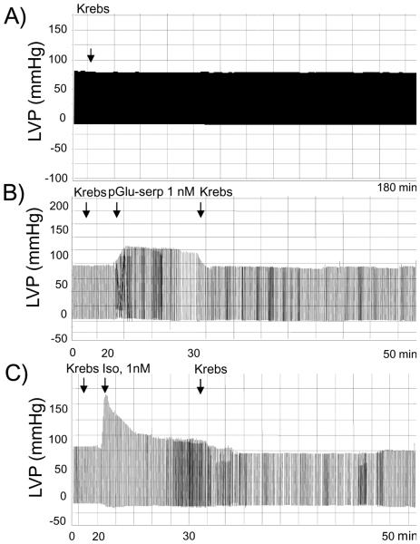
Serpinins are a family of peptides derived from proteolytic cleavage of the penultimate and the last pair of basic residues at the C-terminus of Chromogranin A. Three forms of naturally occurring serpinin have been found in AtT-20 pituitary cells and rat heart. They are serpinin, pyrogutaminated (pGlu) -serpinin and a C-terminally extended form, serpinin-RRG. In addition pGlu-serpinin has been found in brain, primarily in neurites and nerve terminals and shown to have protective effects against oxidative stress on neurons and pituitary cells. Serpinin has also been demonstrated to regulate granule biogenesis in endocrine cells by up-regulating the protease inhibitor, protease nexin-1 transcription via a cAMP-PKA-sp1 pathway. This leads to inhibition of granule protein degradation in the Golgi complex which in turn promotes granule formation. More recently, pGlu-serpinin has been demonstrated to enhance both myocardial contractility (inotropy) and relaxation (lusitropy). In the Langendorff perfused rat heart, pGlu-serpinin showed a concentration-dependent positive inotropic effect exerted through a cAMP-PKA dependent pathway. In conclusion, the serpinin peptides have profound effects at many levels that affect the endocrine and nervous systems and cardiac function.


Figures

Fig. (1A)
Fig. (1A)
Schematic diagram of CgA structure showing the paired basic residues which are potential processing sites, and the serpinin peptide domain. (B) Schematic showing a predicted pGlu-serpinin biosynthetic pathway from mouse CgA based on the endogenous serpinin-related peptides identified. Ion mass of pGlu-serpinin (m/z 2532.5), pGlu-serpinin precursor 1 (m/z 2664.4), serpinin (m/z 2864.5) and serpinin precursor (m/z 3020.6) identified by MALDI-TOF analysis. pGlu-serpinin precursor 2 is a predicted intermediate of pGlu-serpinin biosynthesis.
Fig. (2)
Fig. (2)
Secretion of serpinin and pGlu-serpinin from AtT-20 cells. (A) The <3 kDa fraction of conditioned media from 6T3-bCgA cells and 6T3-WT cells were analyzed for serpinin by EIA. Immunoreactive serpinin was detected in 6T3-bCgA cells, while no immunoreactive serpinin was detected in 6T3-WT cells (n = 4). (B) AtT-20 cells were incubated with high (50 mM) K+ (HK) or low K+ (5 mM) medium (Basal) for 20 min and the <3 kDa fraction of the media assayed by EIA. The amount of immunoreactive serpinin detected in HK-stimulated cell medium was significantly higher than that in basal medium (n = 7, Student’s t-test; *P < 0.05). (C) Using a pGlu-serpinin-specific ELISA, immunoreactive pGlu-serpinin was detected in AtT-20 cell conditioned media, but none was detected in mock (medium alone without cells), or conditioned medium from 6T3 cells that do not express CgA (n = 3). (D) Bar graphs show pGlu-serpinin in basal (2 hr and 10 min) and high K+ stimulation (10 min) incubation media from AtT-20 cells (n = 2).
Fig. (3)
Fig. (3)
Schematic of serpinin/pGlu-serpinin formation, its signaling pathway and PN-1-dependent biogenesis of secretory granules in (neuro)endocrine cells. CgA is proteolytically cleaved to form serpinin which is secreted in an activity-dependent manner. Secreted serpinin/pGlu-serpinin binds to a cognate receptor and induces cAMP elevation followed by PKA activation. Then, Sp1, a transcriptional factor translocates into the nucleus to up-regulate PN-1 transcription. The increase in PN-1 protein stabilizes the secretory granule proteins at the Golgi apparatus to increase their levels which then promotes biogenesis of dense core granules (DCG).
Fig. (4)
Fig. (4)
Serpinin and pGlu-serpinin up-regulated PN-1 mRNA expression. Bar graphs show the effect of serpinin (n = 4) and pGlu-serpinin (n = 3) on PN-1 mRNA expression after treatment of AtT-20 cells for 20 hr with these peptides. Serpinin and pGlu-serpinin significantly up-regulated the PN-1 mRNA at 10 nM (*P < 0.05) and 10 pM (*P < 0.05) concentrations, respectively, relative to untreated cells (Con., n = 3). [From JOMN 2011, 49:294-303].
Fig. (5)
Fig. (5)
Anti-apoptotic effects of pGlu-serpinin and serpinin. (A). AtT-20 cells were challenged with 50 µM hydrogen peroxide in the presence or absence of 1, 10, 100 nM serpinin or 0.1, 1 nM pGlu-serpinin for 1 day. Cell viability was quantified by the LDH assay. Serpinin and pGlu-serpinin significantly inhibited hydrogen peroxide-induced cell death (n = 3, *P < 0.05). (B). Cultured rat cortical neurons were challenged with 50 µM hydrogen peroxide in the presence or absence of 10 nM pGlu-serpinin for 1 day. MAP-2 immunosignal intensity was quantified. pGlu-serpinin significantly inhibited hydrogen peroxide-induced neuronal cell death (n = 3 view fields, P < 0.05; N.S., no significant difference). [From JOMN 2011, 49:294-303].
Fig. (6)
Fig. (6)
Representative LVP (basal value = 89 ± 3 mm Hg) traces showing (A) the time course obtained in the presence of vehicle alone (Krebs), (B) the effect of pGlu-serpinin alone (1 nM), (C) the effects of ISO alone (1 nM) (each arrow represents the administration of a single concentration of the substance or vehicle).

References

    1. Helle KB, Corti A, Metz-Boutigue MH, Tota B. The endocrine role for chromogranin A: a prohormone for peptides with regulatory properties. Cell Mol Life Sci. 2007;64(22):2863–86. - PMC - PubMed
    1. Bartolomucci A, Possenti R, Mahata SK, Fischer-Colbrie R, Loh YP, Salton SRK. The Extended Granin Family: Structure, Function and Biomedical Implications. Endocrine Reviews. 2011;32(6):755–97. - PMC - PubMed
    1. Wohlfarter T, Fischer-Colbrie R, Hogue-Angeletti R, Eiden LE, Winkler H. Processing of chromogranin A within chromaffin granules starts at C- and N-terminal cleavage sites. FEBS Lett. 1988;231(1):67–70. - PubMed
    1. Hook V, Bark S, Gupta N, Lortie M, Lu WD, Bandeira N, Funkelstein L, Wegrzyn J, O'Connor DT, Pevzner P. Neuropeptidomic components generated by proteomic functions in secretory vesicles for cell-cell communication. AAPS J. 2010;12(4):635–45. - PMC - PubMed
    1. Steiner DF. The proprotein convertases. Curr Opin Chem Biol. 1998;2(1):31–9. - PubMed

Publication types

MeSH terms