Review
. 2013 Jan;13(1):24-57.

Functions of S100 proteins

Affiliations
Review

Functions of S100 proteins

R Donato et al. Curr Mol Med. 2013 Jan.

Abstract

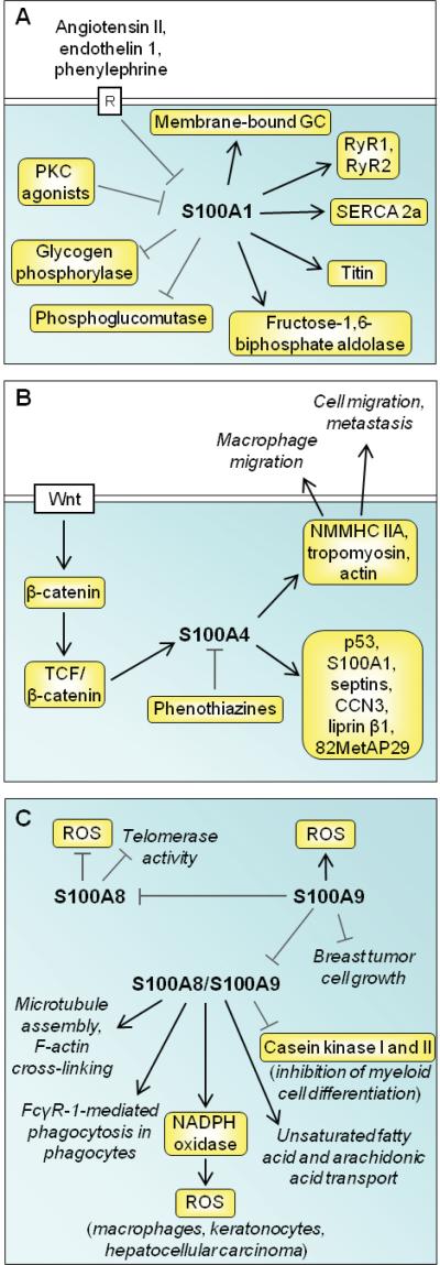
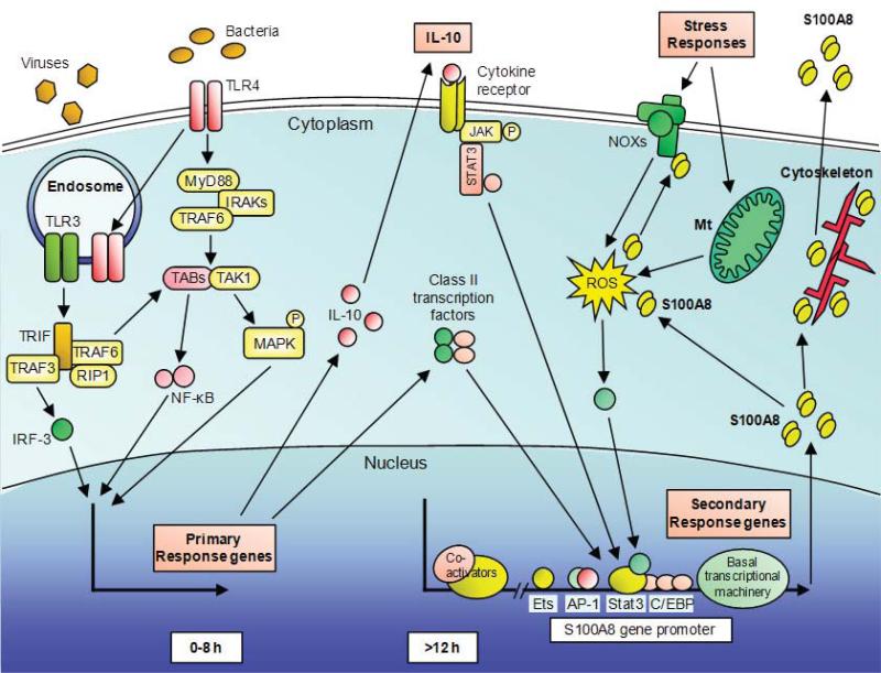
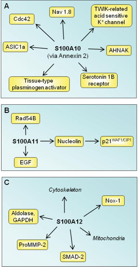
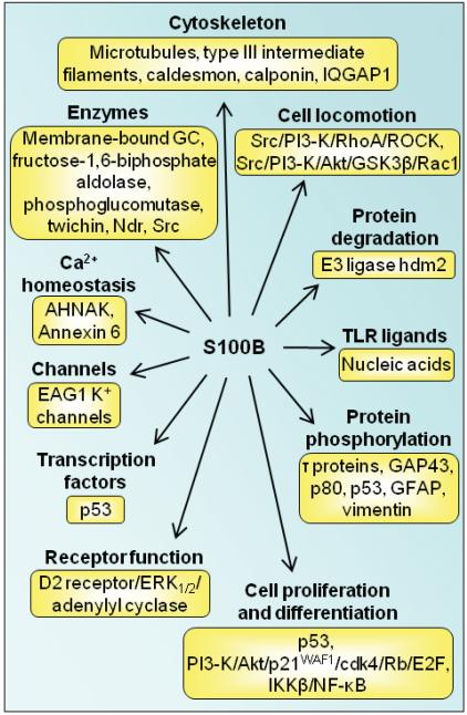
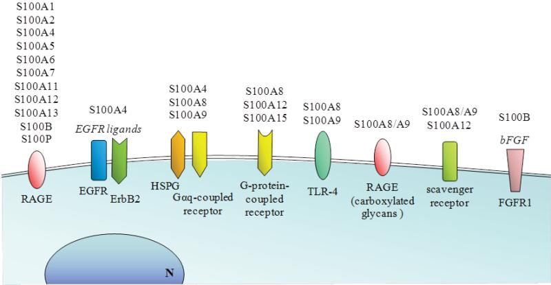
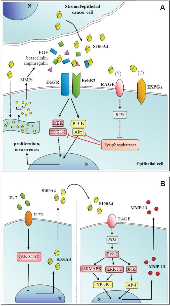
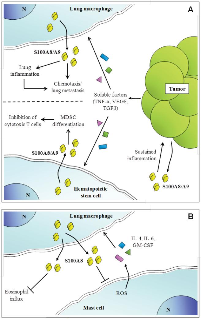
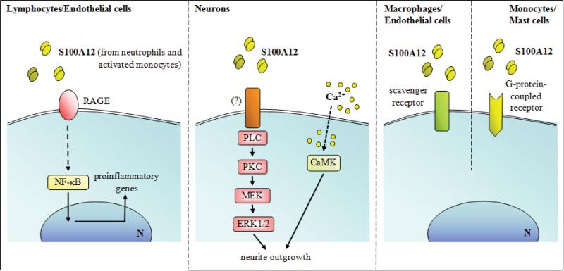
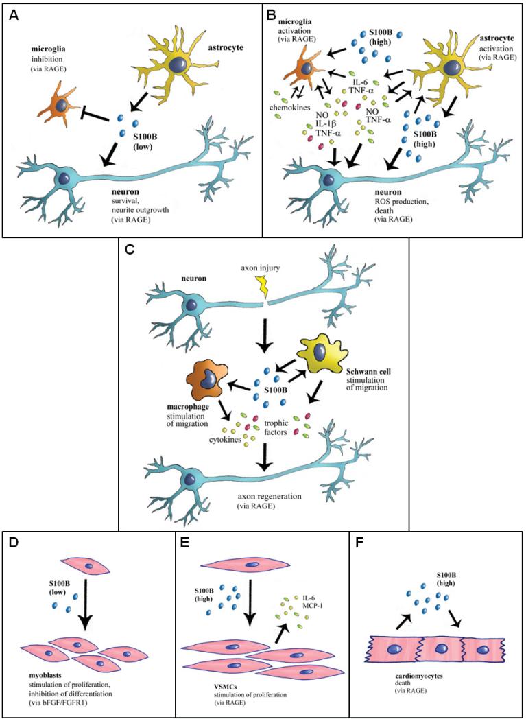
The S100 protein family consists of 24 members functionally distributed into three main subgroups: those that only exert intracellular regulatory effects, those with intracellular and extracellular functions and those which mainly exert extracellular regulatory effects. S100 proteins are only expressed in vertebrates and show cell-specific expression patterns. In some instances, a particular S100 protein can be induced in pathological circumstances in a cell type that does not express it in normal physiological conditions. Within cells, S100 proteins are involved in aspects of regulation of proliferation, differentiation, apoptosis, Ca2+ homeostasis, energy metabolism, inflammation and migration/invasion through interactions with a variety of target proteins including enzymes, cytoskeletal subunits, receptors, transcription factors and nucleic acids. Some S100 proteins are secreted or released and regulate cell functions in an autocrine and paracrine manner via activation of surface receptors (e.g. the receptor for advanced glycation end-products and toll-like receptor 4), G-protein-coupled receptors, scavenger receptors, or heparan sulfate proteoglycans and N-glycans. Extracellular S100A4 and S100B also interact with epidermal growth factor and basic fibroblast growth factor, respectively, thereby enhancing the activity of the corresponding receptors. Thus, extracellular S100 proteins exert regulatory activities on monocytes/macrophages/microglia, neutrophils, lymphocytes, mast cells, articular chondrocytes, endothelial and vascular smooth muscle cells, neurons, astrocytes, Schwann cells, epithelial cells, myoblasts and cardiomyocytes, thereby participating in innate and adaptive immune responses, cell migration and chemotaxis, tissue development and repair, and leukocyte and tumor cell invasion.


Figures

Fig. (1)
Fig. (1)
Coordinating residues for the canonical (site 2) and S100 EF-hand (site 1) for the S100 protein, S100B. In the table below the residues typically at each position of the EF-hand are illustrated. It should be noted that the S100 EF-hand has 14 rather than 12 residues.
Fig. (2)
Fig. (2)
Ribbon diagram illustrating the rotation of helix 3 upon the addition of calcium and a table listing the degree of movement upon calcium and target protein and/or drug binding. The dissociation constants are from [8].
Fig. (3)
Fig. (3)
Schematic representation of proposed intracellular effects of S100A1, S100A4, and S100A8/S100A9. (A) S100A1 expression is negatively controlled by transcription factors downstream of G-protein-coupled receptors and PKC. S100A1 regulates energy metabolism and Ca2+ efflux from Ca2+ stores, stimulates striated muscle contraction, and activates a membrane-bound form of guanylate cyclase (GC) in photoreceptors in relation to dark adaptation. (B) S100A4 is induced by a Wnt/APC/GSK3/β-catenin/TCF pathway and targets several intracellular factors including NMMHC IIA, tropomyosin and actin with ensuing stimulation of cell migration and metastasis. Phenothiazines blocks intracellular S100A4 interactions. (C) S100A8 reduces telomerase activity and ROS production under the negative control of S100A9. S100A9 promotes ROS production, reduces breast cancer cell growth and negatively regulates S100A8/S100A9 heterotetramer complex activities as shown.
Fig. (4)
Fig. (4)
Schematic representation of S100A8 induction in macrophages. LPS from bacteria is recognized by the surface receptor TLR4, activating MyD88-dependent and independent pathways. IRAKs and TRAF6 are recruited to MyD88 and subsequently activates a complex of TAK1 and TABs resulting phosphorylation of IκB and nuclear translocation of NF-κB. Simultaneously, TAK1 activates MAP kinase cascades leading to activation of AP-1. For the MyD88-independent pathway, TLR4 translocates to the endosome together with TRAM. In addition, TLR3 in the endosome, recognizes viral dsRNA. TLR3 and TLR4 activate TRIF-dependent signaling and subsequently activate NF-κB and IRF3. TLR-3 and TLR-4 activation triggers S100A8 gene induction in macrophages, but requires other factors. Induction is a late event that relies on de novo synthesized proteins, particularly IL-10, and class II transcription factors e.g. C/EBPs. AP-1 and Stat-3 bind to the S100A8 promoter. S100A8 is considered a stress response gene, and intracellular ROS generation either via NOXs or mitochondria (Mt) may be essential for induction. Intracellular S100A8, together with S100A9, can interact with components of the cytoskeleton and may mediate their rearrangements and dynamics. S100A8 and S100A9 directly bind to components of the NOX complex and mediate its activity. On the other hand, S100A8 is a potent oxidant scavenger and oxidative modifications of S100A8 can change its functions. S100A8 is actively secreted via a non-classical pathway which requires a functional microtubule network to exert its extracellular functions.
Fig. (5)
Fig. (5)
Schematic representation of proposed intracellular effects of S100A10, S100A11 and S100A12. (A) S100A10 is implicated in the mechanism of action of antidepressant drugs via interaction with serotonin 1B receptor. By binding annexin 2, S100A10 assists the traffic of several membrane proteins to plasma membranes. (B) S100A11 participates in the regulation of cell cycle by several mechanism as shown. (C) S100A12 regulates cytoskeleton-membrane interactions and has Ca2+-dependent chaperone/anti-chaperone-like functions.
Fig. (6)
Fig. (6)
Schematic representation of proposed intracellular effects of S100B. S100B interacts with several intracellular proteins as shown thereby regulating protein phosphorylation, enzyme activities, the state of assembly of certain cytoskeleton components, the transcription factor p53, protein degradation, cell proliferation, locomotion and differentiation, dark adaptation of photoreceptors, Ca2+ homeostasis and the innate inflammatory response.
Fig. (7)
Fig. (7)
Schematic representation of the receptors involved in the transduction of S100 protein signaling. RAGE is an established receptor for several S100 protein members in a variety of cell types. By interacting with EGFR ligands and bFGF, S100A4 and S100B can also activate EGFR and FGFR1, respectively. By interacting with heparan sulfate proteoglycans (HSPG) S100A4, S100A8 and S100A9 also activate Gαq receptors. S100A8, S100A12 and S100A15 can activate G-protein-coupled receptors. S100A8 and/or and S100A9 interact with TLR-4 in phagocytes, and as a heterocomplex S100A8/S100A9 can bind to carboxymethylated RAGE. S100A8/S100A9 and S100A12 also activate scavenger receptors.
Fig. (8)
Fig. (8)
Schematic representation of proposed extracellular effects of S100A4 on epithelial tumor cells (A) and articular chondrocytes (B).
Fig. (9)
Fig. (9)
Schematic representation of proposed extracellular effects of S100A8/S100A9. (A) Activated by TNF-α, TGF-β and VEGF secreted by distant tumors, lung macrophages release S100A8/S100A9 that promotes local inflammation and attract metastatic cells thus promoting tumor cell homing in the lung. Moreover, S100A8/S100A9 might sustain inflammation in the original tumor site. (B) It is also suggested that S100A8/S100A9 released by hematopoietic stem cells under the action of TNF-α, TGF-β and VEGF might inhibit cytotoxic T cells thereby contributing to tumor growth by several mechanisms.
Fig. (10)
Fig. (10)
Schematic representation of proposed extracellular effects of S100A12 on lymphocytes, endothelial cells, neurons, and macrophages.
Fig. (11)
Fig. (11)
Schematic representation of proposed extracellular effects of S100B on neurons microglia, astrocytes, myoblasts, VSMCs, cardiomyocytes, and peripheral nerves. (A) In normal physiological conditions S100B secreted by astrocytes exerts trophic effects on neurons and modulates microglial activity by engaging RAGE. (B) When present in the brain extracellular space at high concentrations, S100B activates microglia and astrocytes thus participating in the inflammatory response and is toxic to neurons by excessively stimulating RAGE. (C) In acute peripheral nerve injury S100B released from activated Schwann cells promotes macrophages and Schwann cell migration and the release of trophic factors via RAGE engagement thereby participating in peripheral nerve regeneration. (D) At subnanomolar-nanomolar doses S100B stimulates myoblast proliferation and inhibits myoblast differentiation by enhancing bFGF/FGFR1 signaling. However, in low-density myoblast cultures and at the very beginning of skeletal muscle regeneration, S100B engages RAGE in activated muscle satellite cells thereby stimulating proliferation and activating the myogenic program. (E) At high doses S100B stimulates VSMC proliferation and secretion of IL-6 and MCP-1 from VSMCs via RAGE engagement thus potentially participating in atherogenesis. (F) At high doses S100B causes cardiomyocyte apoptosis via RAGE engagement.

References

    1. Carafoli E, Klee C. Calcium as a Cellular Regulator. Oxford University Press; New York: 1999.
    1. Permyakov EA, Kretsinger RH. Cell signaling, beyond cytosolic calcium in eukaryotes. J Inorg Biochem. 2009;103:77–86. - PubMed
    1. Donato R. S100: a multigenic family of calcium-modulated proteins of the EF-hand type with intracellular and extracellular functional roles. Int J Biochem Cell Biol. 2001;2001;33:637–8. - PubMed
    1. Marenholz I, Heizmann CW, Fritz G. S100 proteins in mouse and man: from evolution to function and pathology (including an update of the nomenclature). Biochem Biophys Res Commun. 2004;322:1111–22. - PubMed
    1. Donato R. RAGE: a single receptor for several ligands and different cellular responses: the case of certain S100 proteins. Curr Mol Med. 2007;7:711–24. - PubMed

Publication types