Review
. 2012 May;32(3):295-303.
doi: 10.1016/j.semnephrol.2012.04.009.

Transforming growth factor-β, bioenergetics, and mitochondria in renal disease

Affiliations
Review

Transforming growth factor-β, bioenergetics, and mitochondria in renal disease

Gabriella Casalena et al. Semin Nephrol. 2012 May.

Abstract

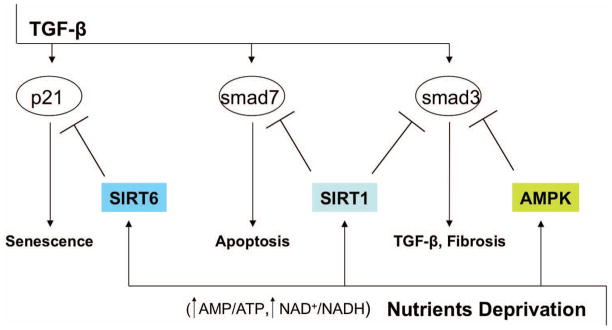
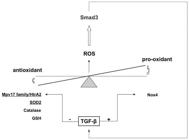
The transforming growth factor-β (TGF-β) family comprises more than 30 family members that are structurally related secreted dimeric cytokines, including TGF-β, activins, and bone morphogenetic proteins/growth and differentiation factors. TGF-β are pluripotent regulators of cell proliferation, differentiation, apoptosis, migration, and adhesion of many different cell types. TGF-β pathways are highly evolutionarily conserved and control embryogenesis, tissue repair, and tissue homeostasis in invertebrates and vertebrates. Aberrations in TGF-β activity and signaling underlie a broad spectrum of developmental disorders and major pathologies in human beings, including cancer, fibrosis, and autoimmune diseases. Recent observations have indicated an emerging role for TGF-β in the regulation of mitochondrial bioenergetics and oxidative stress responses characteristic of chronic degenerative diseases and aging. Conversely, energy and metabolic sensory pathways cross-regulate mediators of TGF-β signaling. Here, we review TGF-β and regulation of bioenergetic and mitochondrial functions, including energy and oxidant metabolism and apoptotic cell death, as well as their emerging relevance in renal biology and disease.


Figures

Figure 1
Figure 1
TGF-β targets in mitochondria. TGF-β signaling may act in a direct or indirect manner to control mitochondrial metabolism in different cell types. ⍊ and ↓ indicate inhibition/downregulation by TGF-β pathway. Oxidative phosphorylation system (I-V), membrane potential (ΔΨ), reactive oxygen species (ROS), endoplasmic reticulum (ER). Inhibition of the oxidative phosphorylation system may arise from a direct inhibition of complex IV and/or from increased ROS production due to downregulation of scavenging proteins (MnSOD, Mpv17l). Decrease in membrane potential is associated with cytochrome c release in the cytoplasm with consequent activation of the apoptotic signaling. Apoptosis can be triggered by release of HtrA2 as a consequence of Mpv17l downregulation. Uncoupling of mitochondria from ER calcium release may impair the intracellular calcium sensing responses.
Figure 2
Figure 2
TGF-β and AMPK-SIRT crosstalk. The negative cross-regulation between TGF-β signaling and energy sensing mediators AMPK and SIRT has been demonstrated in kidney, liver, and lung. AMPK and SIRTs may downregulate TGF-β messengers either by degradation or inhibition of transcriptional activity. AMPK mediates inhibition of transcription co-activator p300 and Smad3 interaction in hepatic stellate cells. SIRT1-mediated deacetylation of Smad3 and Smad7 results in reduction of apoptosis and fibrosis in kidney cells in vitro and in vivo. SIRT6-mediated proteasomal degradation of p21 prevents TGF-β-induced senescence in primary bronchial epithelial cells.
Figure 3
Figure 3
TGF-β and oxidative stress. TGF-β increases oxidative stress by regulating both antioxidant and pro-oxidant proteins. Downregulation of mitochondrial (underline) Mpv17-HtrA2 system, manganese superoxide-dismutase (SOD2), catalase, and glutathione (GSH) with the concomitant stimulation of pro-oxidant NADPH-oxidase (Nox4) results in increased ROS production that may trigger a positive-feedback response in which TGF-β signaling is amplified by Smad3.

References

    1. Xue JL, Ma JZ, Louis TA, Collins AJ. Forecast of the number of patients with end-stage renal disease in the United States to the year 2010. J Am Soc Nephrol. 2001;12:2753–8. - PubMed
    1. Collins AJ, Foley RN, Herzog C, et al. Excerpts from the US Renal Data System 2009 Annual Data Report. Am J Kidney Dis. 55:S1–420. A6–7. - PMC - PubMed
    1. Bottinger EP. TGF-beta in renal injury and disease 4. Semin Nephrol. 2007;27:309–20. - PubMed
    1. Kagami S, Border WA, Miller DE, Noble NA. Angiotensin II stimulates extracellular matrix protein synthesis through induction of transforming growth factor-beta expression in rat glomerular mesangial cells. J Clin Invest. 1994;93:2431–7. - PMC - PubMed
    1. Okuda S, Languino LR, Ruoslahti E, Border WA. Elevated expression of transforming growth factor-beta and proteoglycan production in experimental glomerulonephritis. Possible role in expansion of the mesangial extracellular matrix [published erratum appears in J Clin Invest 1990 Dec;86(6):2175] J Clin Invest. 1990;86:453–62. - PMC - PubMed

Publication types