. 2012 Nov 5;363(1-2):46-61.
doi: 10.1016/j.mce.2012.07.006. Epub 2012 Jul 24.

Relaxin induces matrix-metalloproteinases-9 and -13 via RXFP1: induction of MMP-9 involves the PI3K, ERK, Akt and PKC-ζ pathways

Affiliations

Relaxin induces matrix-metalloproteinases-9 and -13 via RXFP1: induction of MMP-9 involves the PI3K, ERK, Akt and PKC-ζ pathways

Nisar Ahmad et al. Mol Cell Endocrinol. .

Abstract

We determined the precise role of relaxin family peptide (RXFP) receptors-1 and -2 in the regulation of MMP-9 and -13 by relaxin, and delineated the signaling cascade that contributes to relaxin's modulation of MMP-9 in fibrocartilaginous cells. Relaxin treatment of cells in which RXFP1 was silenced resulted in diminished induction of MMP-9 and -13 by relaxin, whereas overexpression of RXFP1 potentiated the relaxin-induced expression of these proteinases. Suppression or overexpression of RXFP2 resulted in no changes in the relaxin-induced MMP-9 and -13. Studies using chemical inhibitors and siRNAs to signaling molecules showed that PI3K, Akt, ERK and PKC-ζ and the transcription factors Elk-1, c-fos and, to a lesser extent, NF-κB are involved in relaxin's induction of MMP-9. Our findings provide the first characterization of signaling cascade involved in the regulation of any MMP by relaxin and offer mechanistic insights on how relaxin likely mediates extracellular matrix turnover.


Figures

Fig. 1
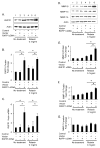
Fig. 1. Overexpression of RXFP1 potentiates relaxin’s induction of MMP-9 and -13 in fibrochondrocytes
Untransfected cells (Control) or cells transiently transfected with pcDNA vector, or vector containing RXFP1 cDNA were cultured in the absence or presence of relaxin (0.1ng/ml). RNA, cell-matrix extract and cell-conditioned medium were collected after 48 hours and assayed. cDNA transfection successfully enhanced RXFP1 protein expression assayed by Western blots (A) and quantitated by videodensitometry (B), as well as mRNA assayed by qRT-PCR (C) in both untreated and relaxin-treated cells. Western blots for MMPs quantitated by videodensitometry revealed that relaxin induced MMP-9 (D and E) and -13 (D and F), but not MMP-14 (D and G). Cells overexpressing RXFP1 showed enhanced induction of MMP-9 (D and E) and -13 (D and F) by relaxin. Quantitative data is shown as mean (±SE) fold-change in RXFP1 or MMP levels relative to control untransfected and untreated cells. Actin was used as a loading control for Western blots. (*p < 0.05).
Fig. 2
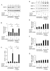
Fig. 2. Overexpression of RXFP2 does not modulate the expression of MMP-9 and -13 in fibrochondrocytes
Untransfected cells (Control) or cells transiently transfected with pcDNA vector, RXFP2 cDNA constructs were cultured in the absence or presence of relaxin (0.1ng/ml). RNA, cell-matrix extract and cell-conditioned medium were collected after 48 hours and assayed. cDNA transfection successfully enhanced RXFP2 protein expression assayed by Western blots (A) and quantitated by videodensitometry (B), as well as mRNA assayed by qRT-PCR (C) in both untreated and relaxin-treated cells. Western blots for MMPs quantitated by videodensitometry revealed that relaxin induced MMP-9 (D and E) and -13 (D and F), but not MMP-14 (D and G). Cells overexpressing RXFP2 showed no further modulation of MMP-9 (D and E) and -13 (D and F) by relaxin. Quantitative data is shown as mean (± SE) fold-change in RXFP2 or MMP levels relative to control untransfected and untreated cells. Actin was used as a loading control for Western blots. (*p < 0.05).
Fig. 3
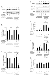
Fig. 3. Inhibition of RXFP1 expression negates the ability of relaxin to induce MMP-9 and -13 in fibrochondrocytes
Untransfected cells (Control) or cells transiently transfected with scrambled siRNA, or siRNA to RXFP1 were cultured in the absence or presence of relaxin (0.1ng/ml). RNA, cell-matrix extract and cell-conditioned medium were collected after 48 hours and assayed. siRNA transfections inhibited RXFP1 protein expression assayed by Western blots (A) and quantitated by videodensitometry (B), as well as mRNA assayed by qRT-PCR (C) in both untreated and relaxin-treated cells. Western blots for MMPs quantitated by videodensitometry revealed that relaxin induced MMP-9 (D and E) and -13 (D and F), but not MMP-14 (D and G). Suppression of RXFP1 decreased both the constitutive expression levels and relaxin’s induction of MMP-9 (D and E) and -13 (D and F). Quantitative data is shown as mean (±SE) fold-change in RXFP1 or MMP levels relative to control untransfected and untreated cells. Actin was used as a loading control for Western blots. (*p < 0.05).
Fig. 4
Fig. 4. Suppression of RXFP2 does not contribute to relaxin’s modulation of MMP-9 and -13
Untransfected cells (Control) or cells transiently transfected with scrambled siRNA, or siRNA to RXFP2 were cultured in the absence or presence of relaxin (0.1ng/ml). RNA, cell-matrix extract and cell-conditioned medium were collected after 48 hours and assayed. siRNA transfections inhibited RXFP2 protein expression assayed by Western blots (A) and quantitated by videodensitometry (B), as well as mRNA assayed by qRT-PCR (C) in both untreated and relaxin-treated cells. Western blots for MMPs quantitated by videodensitometry revealed that relaxin induced MMP-9 (D and E) and -13 (D and F), but not MMP-14 (D and G). Suppression of RXFP2 did not modulate relaxin’s induction of MMP-9 (D and E) and -13 (D and F). Quantitative data is shown as mean (±SE) fold-change in RXFP1 or MMP levels relative to control untransfected and untreated cells. Actin was used as a loading control for Western blots. (*p < 0.05).
Fig. 5
Fig. 5. Relaxin induces MMP -9 and -13 via RXFP1 but not RXFP2
Cells transiently transfected with pcDNA vector or RXFP1 cDNA or RXFP2 cDNA (A and B), or with scrambled siRNA or RXFP1 siRNA or RXFP2 siRNA (C and D), were cultured in the absence or presence of relaxin (0.1ng/ml). Total RNA was isolated, reverse transcribed and subjected to qRT-PCR using MMP-9 (A and C) and MMP-13 (B and D) specific primers. Data is shown as mean (±SE) fold-change in MMP mRNA levels relative to empty vector transfected and untreated cells. (*p < 0.05).
Fig. 6
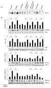
Fig. 6. Activation of RXFP1 by relaxin promotes phosphorylation of signaling molecules PI3K, Akt, PKC-ζ, and ERK1/2 in parallel with induction of MMP-9
Fibrochondrocytes were pre-treated with signaling inhibitors, 2μM LY294002 (PI3K), 1μM chelerythrine chloride (PKC), 2μM PKC-mrystilated pseudosubstrate (PKC-ζ), 1μM tyrphostins (insulin), 1μM U1026 (MEK/ERK) or vehicle (DMSO) for 30 minutes and then stimulated with 0.1ng/ml relaxin. Western blots were performed for MMP-9 in cell-conditioned media collected at 6 hours (A), and for signaling molecules (B) in cell lysates retrieved after 30 minutes. The histograms in B represent mean (± SE) fold change relative to untreated controls of phosphorylated signaling molecule standardized to the respective unphosphorylated protein from Western blots from three independent experiments. Relaxin treatment increased the phosphorylated forms of PI3K, Akt, PKC-ζ and ERK1/2. Relaxin’s induction of MMP-9 was inhibited by PI3K, PKC, PKC-ζ and ERK inhibitors. Tyrphostins, which inhibit insulin signaling, served as negative control. Actin was used a loading control. (*p < 0.05).
Fig. 7
Fig. 7. Relaxin’s induction of MMP-9 expression is Akt dependent
Fibrochondrocytes were transiently transfected with empty vector or PGL4 vector or Akt dominant negative construct (Akt DN) or MMP-9 promoter-luciferase construct and cultured in the absence or presence of relaxin (0.1ng/ml). Cell lysates were collected after 6 hours and subjected to luciferase assay. (A) Relaxin treatment increased the expression of MMP-9-promoter linked luciferase activity, which was significantly reduced in cells transfected with Akt DN construct. (B) Conditioned medium from cells transiently transfected with PGL4 vector and increasing concentrations of Akt DN showed induction of MMP-9 induction by relaxin, which was dose-dependently reduced in the presence of Akt DN construct. (*p<0.05)
Fig. 8
Fig. 8. Activation of RXFP1 by relaxin promotes phosphorylation of transcription factors NF-κB, Elk-1 and c-fos downstream of PI3K, PKC, PKC-ζ and ERK1/2 in parallel with induction of MMP-9
Fibrochondrocytes were pre-treated with signaling inhibitors or vehicle (DMSO) for 30 minutes and then stimulated with 0.1ng/ml relaxin as described in Figure 6. Western blots were performed for MMP-9 in cell-conditioned media collected at 6 hours (A), and transcription factors (B) in cell lysates retrieved after 30 minutes. The histograms in B represent mean (± SE) fold change relative to untreated controls of phosphorylated transcription factors standardized to the respective unphosphorylated proteins from Western blots from three independent experiments. Relaxin treatment increased the phosphorylated forms of NF-κB, Elk-1 and c-fos. Relaxin’s induction of MMP-9 was inhibited by PI3K, PKC, PKC-ζ and ERK inhibitors. Tyrphostins, which inhibit insulin signaling, served as negative control.. Actin was used a loading control. (*p < 0.05).
Fig. 9
Fig. 9. Silencing of ERK1/2, PKC-ζ, Elk-1 eliminates relaxin’s induction of MMP-9
Untransfected cells (Control) or cells transiently transfected with scrambled siRNA, or siRNA to ERK1/2, PKC-ζ, Elk-1 or NF-κB were cultured in the absence or presence of relaxin (0.1ng/ml). Western blots for ERK1/2 (A), PKC-ζ (B), Elk-1 (C) and NF-κB (D) demonstrate successful silencing of the respective molecules and their phosphorylated forms by siRNA transfections. Western blots for MMP-9 show that suppression of ERK1/2, PKC-ζ, or Elk-1 results in elimination of relaxin’s induction of MMP-9 (A to C). (D) NF-κB suppression results in minimal inhibition of induction of MMP-9 by relaxin. The histograms represent mean (± SE) fold change of MMP-9 (white bars) and phosphorylated (black bars) proteins from Western blots from three independent experiments. Actin was used as an internal control. (*p < 0.05).
Fig. 10
Fig. 10. Schematic representation of signaling pathways involved in relaxin’s induction of MMP-9 in fibrochondrocytes
Relaxin binds to RXFP1 receptor and activates PI3K/Akt, ERK and PKC-ζ signaling pathways, which leads to the phosphorylation of DNA binding transcriptional factors Elk-1, c-fos and NF-κB. Silencing of Elk-1 and c-fos and to a lesser extent NF-κB eliminated relaxin-induced MMP-9 expression in fibrochondrocytes.

References

    1. Bani D. Relaxin: a pleiotropic hormone. Gen Pharmacol. 1997;28:13–22. - PubMed
    1. Bathgate RA, Ivell R, Sanborn BM, Sherwood OD, Summers RJ. Receptors for relaxin family peptides. Ann N Y Acad Sci. 2005;1041:61–76. - PubMed
    1. Bathgate RA, Samuel CS, Burazin TC, Gundlach AL, Tregear GW. Relaxin: new peptides, receptors and novel actions. Trends Endocrinol Metab. 2003;14:207–13. - PubMed
    1. Bogatcheva NV, Truong A, Feng S, Engel W, Adham IM, Agoulnik AI. GREAT/LGR8 is the only receptor for insulin-like 3 peptide. Mol Endocrinol. 2003;17:2639–46. - PubMed
    1. Byun HJ, Hong IK, Kim E, Jin YJ, Jeoung DI, Hahn JH, Kim YM, Park SH, Lee H. A splice variant of CD99 increases motility and MMP-9 expression of human breast cancer cells through the AKT-, ERK-, and JNK-dependent AP-1 activation signaling pathways. J Biol Chem. 2006;281:34833–47. - PubMed

Publication types

MeSH terms