Review
. 2012 Sep;144(3):200-13.
doi: 10.1016/j.clim.2012.07.001. Epub 2012 Jul 13.

Metabolic regulation of organelle homeostasis in lupus T cells

Affiliations
Review

Metabolic regulation of organelle homeostasis in lupus T cells

Tiffany N Caza et al. Clin Immunol. 2012 Sep.

Abstract

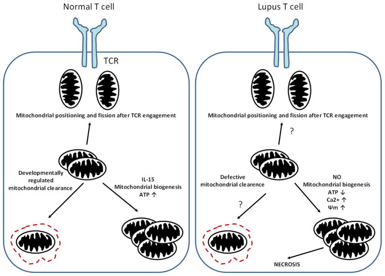
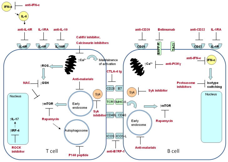
Abnormal T-cell signaling and activation are characteristic features in systemic lupus erythematosus (SLE). Lupus T cells are shifted toward an over-activated state, important signaling pathways are rewired, and signaling molecules are replaced. Disturbances in metabolic and organelle homeostasis, importantly within the mitochondrial, endosomal, and autophagosomal compartments, underlie the changes in signal transduction. Mitochondrial hyperpolarization, enhanced endosomal recycling, and dysregulated autophagy are hallmarks of pathologic organelle homeostasis in SLE. This review is focused on the metabolic checkpoints of endosomal traffic that control immunological synapse formation and mitophagy and may thus serve as targets for treatment in SLE.


Figures

Fig. 1
Fig. 1
Mitochondrial homeostasis in a normal and a lupus T cells. During the formation of the IS, mitochondria are shuttled and redistributed towards the TCR signaling complex. Mitochondrial content is regulated during T cell differentiation allowing damaged mitochondria to be eliminated by autophagy. During T cell memory formation, mitochondrial biogenesis and ATP content are increased via the effect of IL-15. However, in lupus T cell, higher mitochondrial mass and elevated potential is observed, which could be due to higher NO production in these cells. Mitochondria are larger in size and have elevated Ca2+, which alters mitochondrial movement and contributes to defective IS architecture. Removal of mitochondria could be disturbed during T cell differentiation. Contrary to the normal T cell, SLE T cells produce less ATP. The result of mitochondrial dysfunction can be necrotic cell death upon stimulation.
Fig. 2
Fig. 2
Biological targets under investigation for treatment of SLE. Depletion of autoreactive B cells is achieved by treatment with monoclonal antibodies against CD20 and CD22. CD22 also depletes plasma cells, which can also be targeted through blockade of isotype-switching by proteasome inhibition or anti-IFNα antibodies. Activation of lupus B cells can be inhibited by targeting survival (BAFF targeted by Belimumab, APRIL targeted by TACI-Ig) and co-stimulatory signals. Lupus T cell activation is targeted by blockade of cytokine action (IL-6, IL-1, and IL-10), cytokine production (IL-17), and co-stimulation (CD28-B7 interaction by CTLA-4 Ig, ICOS-ICOS ligand interaction by anti-B7RP-1 antibodies). Activation of SLE T and B cells results in a rise of cellular Ca2+, which results from mitochondrial dysfunction, mTOR activation, and endocytic pathway activation. Intracellular Ca2+ can be modulated by treatment with calcium calmodulin kinase inhibitors, calcineurin inhibitors, and anti-PI3Kγ. Early endosome and mTOR activation in SLE T and B cells are inhibited with anti-malarials and rapamycin. Autophagy in SLE lymphocytes can be reduced by treatment with P140 peptide and anti-malarial drugs. Of these targets, antimalarial drugs and Belimumab have been FDA-approved for SLE disease management. The other targets mentioned are under intensive investigation in pre-clinical and clinical studies.

Similar articles

Cited by

References

    1. Tsokos GC. Mechanisms of disease: Systemic lupus erythematosus. N Engl J Med. 2011;365:2110–2121. - PubMed
    1. Edwards CJ, Cooper C. Early environmental exposure and the development of lupus. Lupus. 2006;15:814–819. - PubMed
    1. Moser KL, Kelly JA, Lessard CJ, Harley JB. Recent insights into the genetic basis of systemic lupus erythematosus. Genes Immun. 2009;10:373–379. - PMC - PubMed
    1. Zhou Y, Lu Q. DNA methylation in T cells from idiopathic lupus and drug-induced lupus patients. Autoimmunity Reviews. 2008;7:376–383. - PubMed
    1. Deapen D, Escalante A, Weinrib L, Horwitz D, Bachman B, Roy-Burman P, Walker A, Mack TM. A revised estimate of twin concordance in systemic lupus erythematosus. Arthritis Rheum. 1992;35:311–318. - PubMed