. 2012 Oct;237(2):355-62.
doi: 10.1016/j.expneurol.2012.07.008. Epub 2012 Jul 24.

Low nigrostriatal reserve for motor parkinsonism in nonhuman primates

Affiliations

Low nigrostriatal reserve for motor parkinsonism in nonhuman primates

Samer D Tabbal et al. Exp Neurol. 2012 Oct.

Abstract

Objective: Nigrostriatal reserve refers to the threshold of neuronal injury to dopaminergic cell bodies and their terminal fields required to produce parkinsonian motor deficits. Inferential studies have estimated striatal dopamine reserve to be at least 70%. Knowledge of this threshold is critical for planning interventions to prevent symptom onset or reverse nigrostriatal injury sufficient to restore function in people with Parkinson disease. In this study, we determine the nigrostriatal reserve in a non-human primate model that mimics the motor manifestations of Parkinson disease.

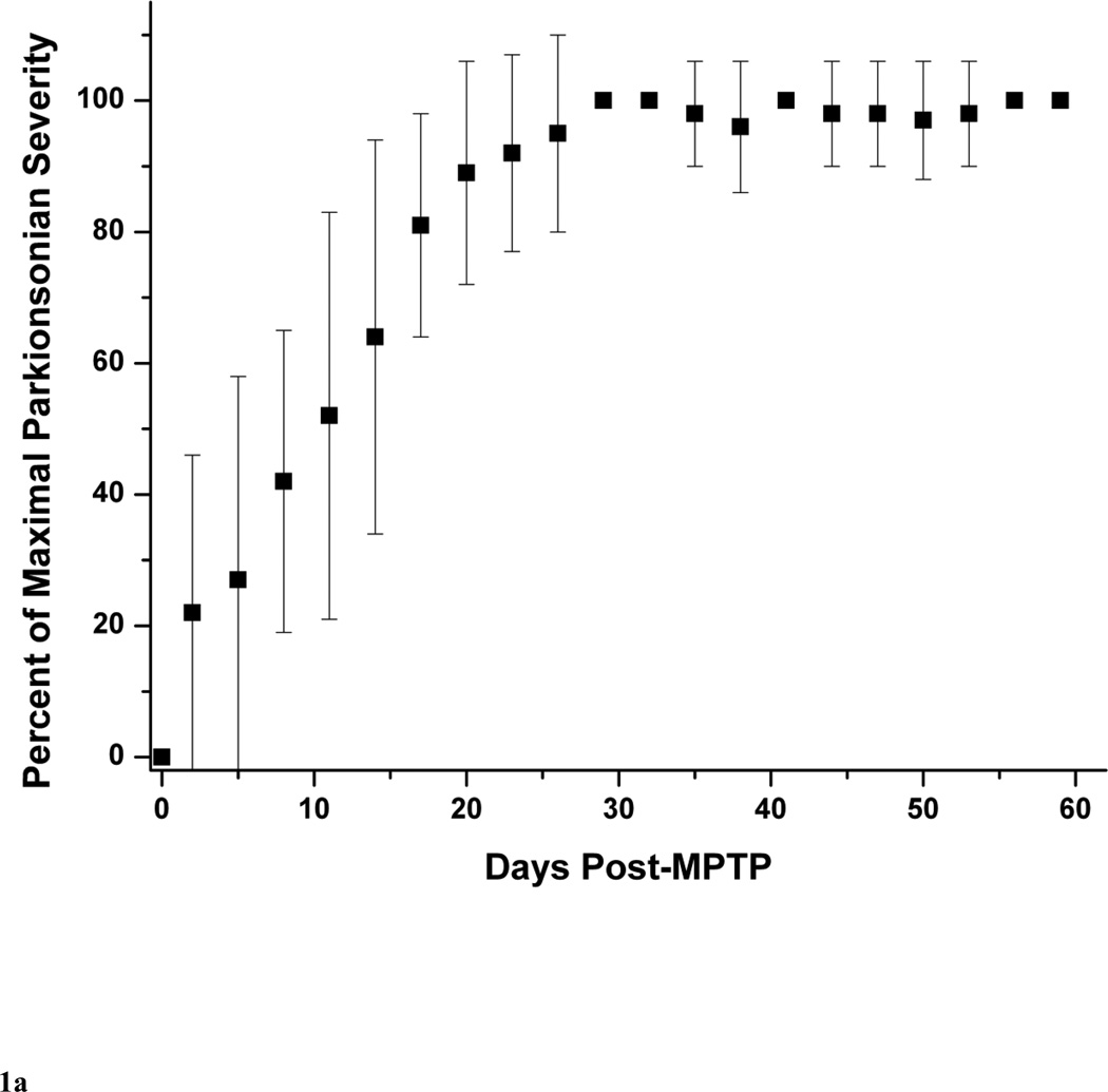
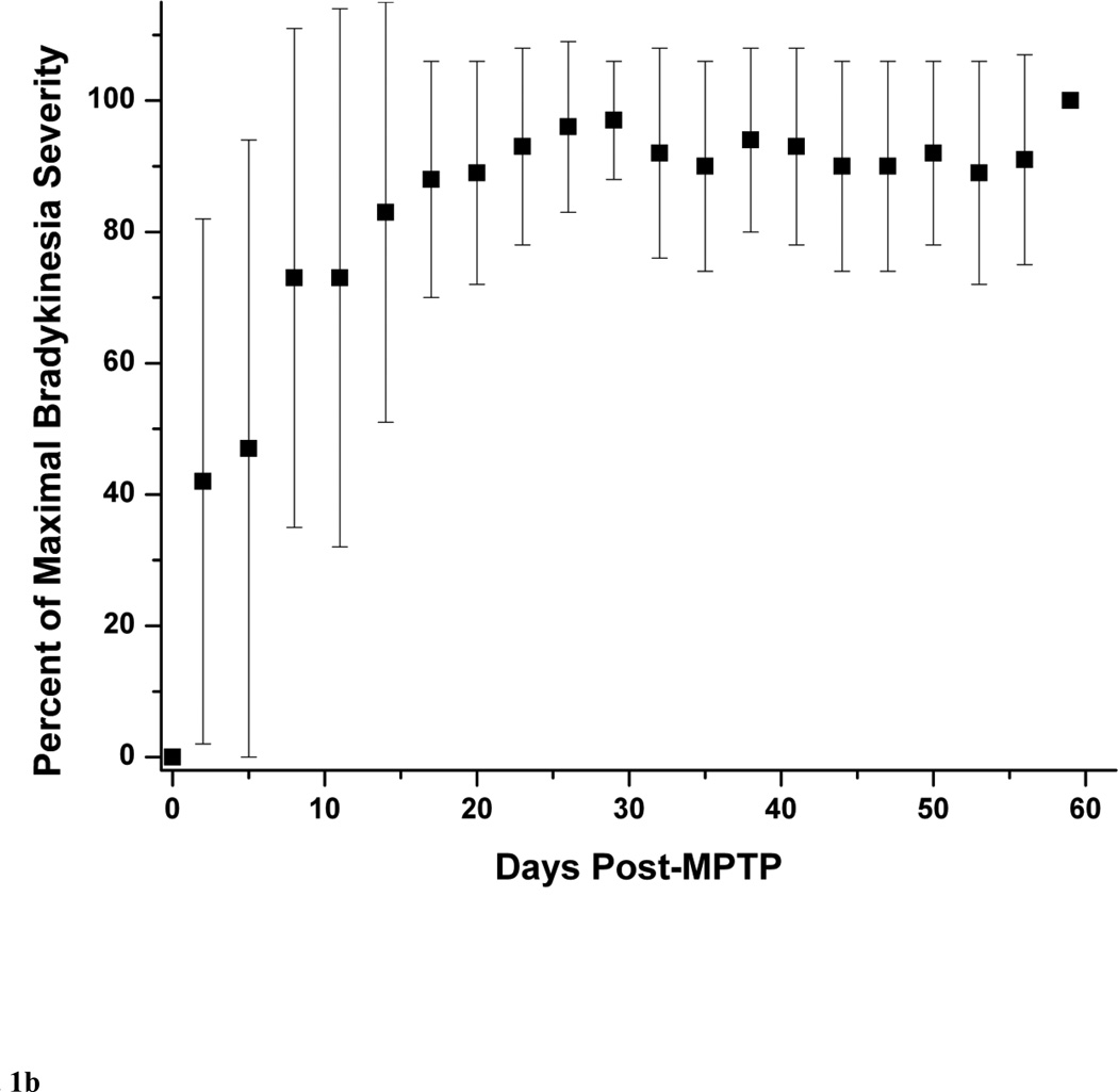
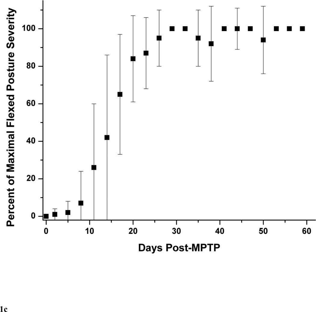
Methods: Fifteen macaque monkeys received unilateral randomized doses of the selective dopaminergic neuronal toxin 1-methyl-4-phenyl-1,2,3,6-tetrahydropyridine. We compared blinded validated ratings of parkinsonism to in vitro measures of striatal dopamine and unbiased stereologic counts of nigral neurons after tyrosine hydroxylase immunostaining.

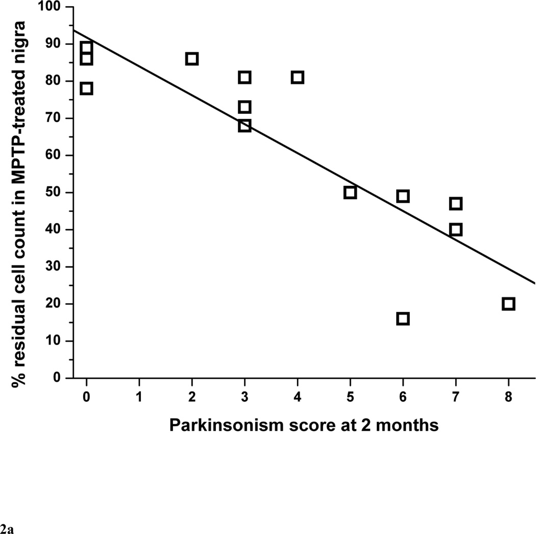
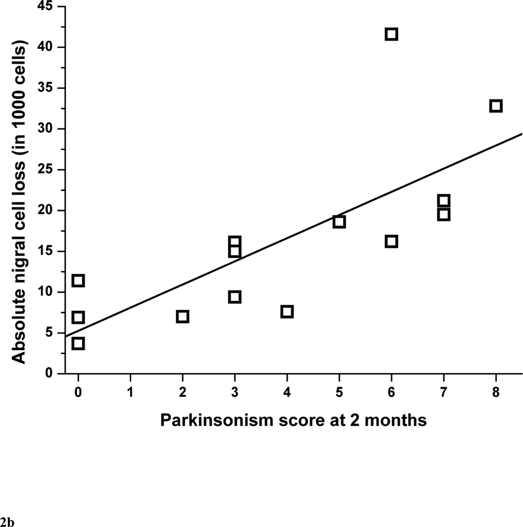
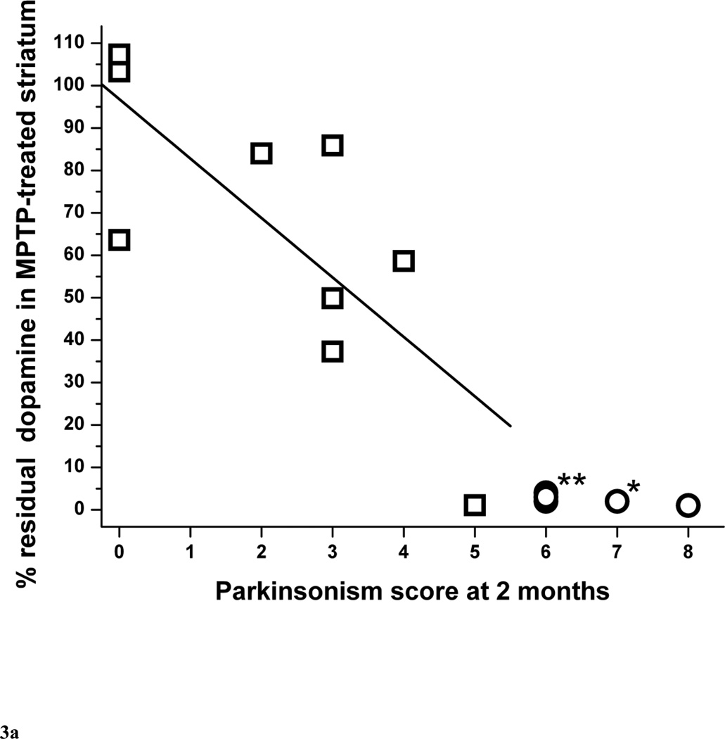
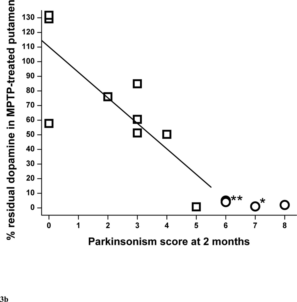
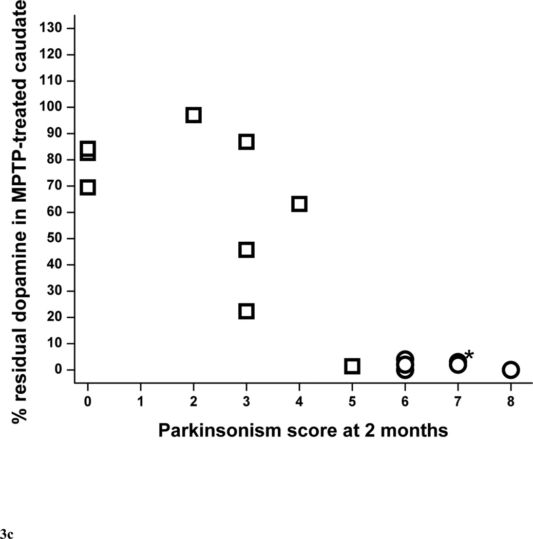
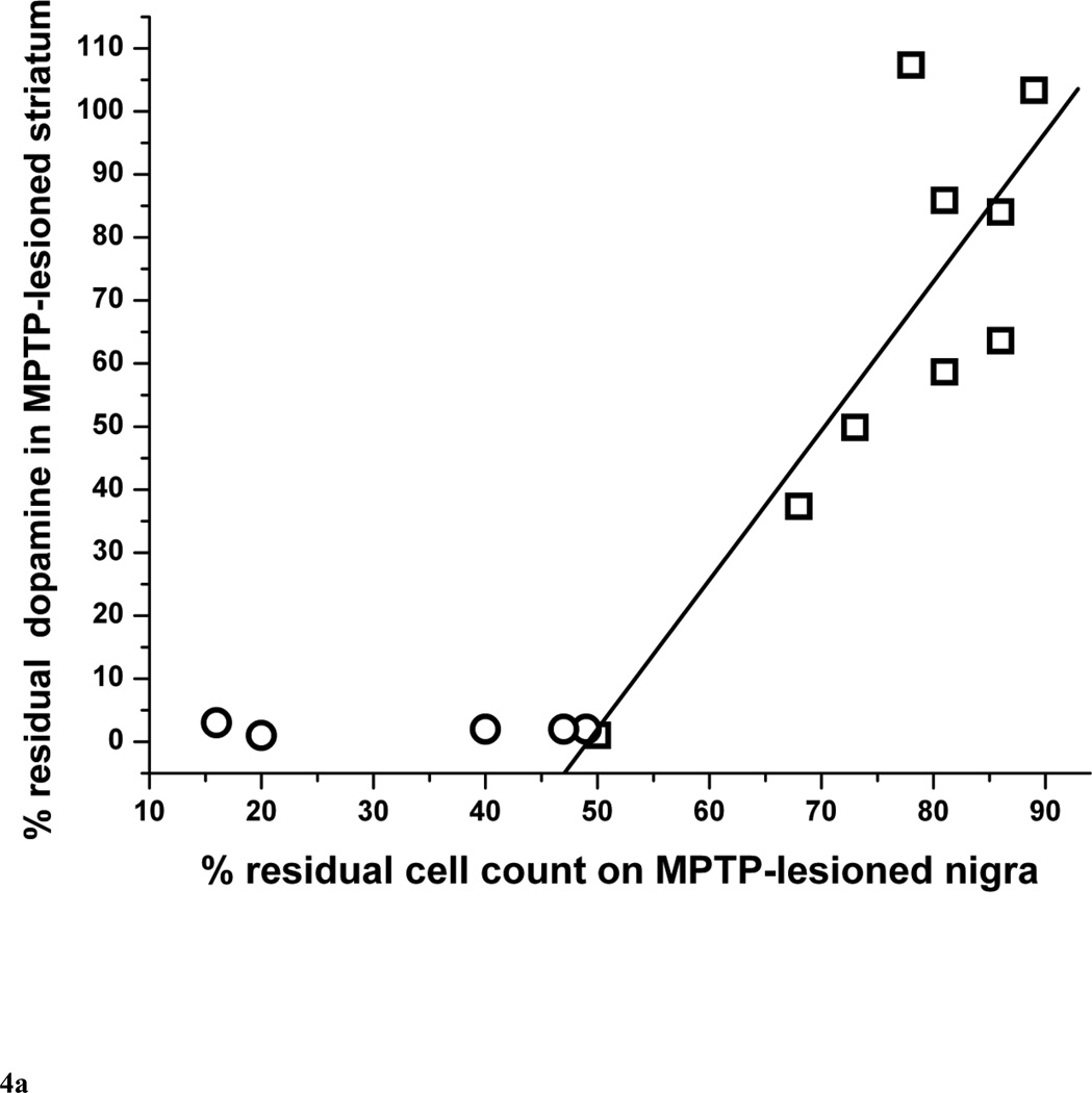
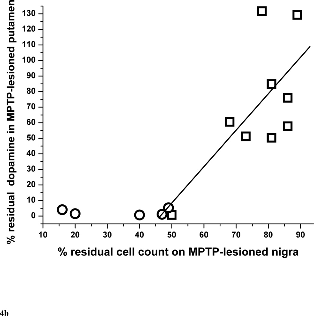
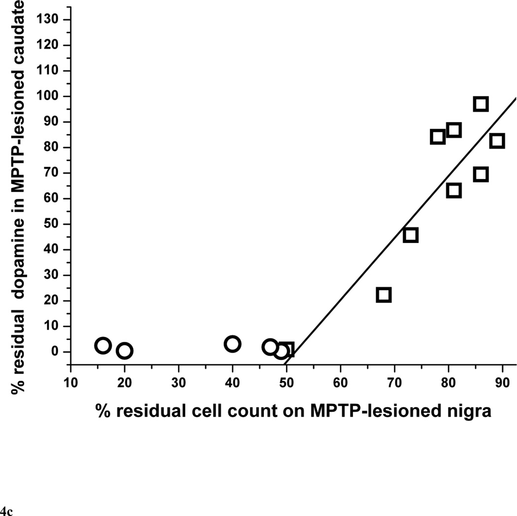
Results: The percent of residual cell counts in lesioned nigra correlated linearly with the parkinsonism score at 2 months (r=-0.87, p<0.0001). The parkinsonism score at 2 months correlated linearly with the percent residual striatal dopamine (r=-0.77, p=0.016) followed by a flooring effect once nigral cell loss exceeded 50%. A reduction of about 14 to 23% of nigral neuron counts or 14% to 37% of striatal dopamine was sufficient to induce mild parkinsonism.

Conclusions: The nigral cell body and terminal field injury needed to produce parkinsonian motor manifestations may be much less than previously thought.


Figures

Figure 1
Figure 1
Behavioral response over 2 months after unilateral intra-carotid MPTP administration, including (A) total parkinsonian score, (B) bradykinesia subscore and (C) flexed posture subscore. Each data point is an average of percent of maximal parkinsonian score severity calculated from several monkeys within a 3-day interval. Percent of maximal parkinsonian score severity for each monkey was defined as the average of parkinsonian score over 3 days divided by the maximal parkinsonian score of that monkey over the course of the study. The error bars represent standard deviations.
Figure 1
Figure 1
Behavioral response over 2 months after unilateral intra-carotid MPTP administration, including (A) total parkinsonian score, (B) bradykinesia subscore and (C) flexed posture subscore. Each data point is an average of percent of maximal parkinsonian score severity calculated from several monkeys within a 3-day interval. Percent of maximal parkinsonian score severity for each monkey was defined as the average of parkinsonian score over 3 days divided by the maximal parkinsonian score of that monkey over the course of the study. The error bars represent standard deviations.
Figure 2
Figure 2
Parkinsonism score at 2 months versus (A) percent of residual dopaminergic cell counts and (B) absolute number of nigral cell loss in MPTP-lesioned nigra (n=14). In figure 2A, the linear equation y = A + B*x has A = 91.8±6.1, B = −7.8±1.3, r = −0.87, p <0.0001. In figure 2B, the linear equation y = A + B*x has A = 5.3±3.5, B = 2.8±0.7, r = 0.74, p = 0.0024.
Figure 2
Figure 2
Parkinsonism score at 2 months versus (A) percent of residual dopaminergic cell counts and (B) absolute number of nigral cell loss in MPTP-lesioned nigra (n=14). In figure 2A, the linear equation y = A + B*x has A = 91.8±6.1, B = −7.8±1.3, r = −0.87, p <0.0001. In figure 2B, the linear equation y = A + B*x has A = 5.3±3.5, B = 2.8±0.7, r = 0.74, p = 0.0024.
Figure 3
Figure 3
Parkinsonism score at 2 months versus percent residual dopamine content in (A) the striatum, (B) putamen and (C) caudate (n=15). Data from monkeys with residual nigral cell counts of less than 50% are depicted by circles (n=6). Squares represent data from monkeys with residual nigral cell counts of 50% or more (n=9). In figure 3A, the linear equation y = A + B*x has A = 96.8±12.5, B = −14.0±4.4, r = −0.77, p = 0.016. In figure 3B, the linear equation has A = 110±14, B = −17.5±5.1, r = −0.79, p = 0.011). In figure 3C, the linear equation has A = 86.4±14.2, B = −11.2±5.0, r = −0.65, p = 0.060. * denotes 2 overlapping data points; ** denotes 3 overlapping data points.
Figure 3
Figure 3
Parkinsonism score at 2 months versus percent residual dopamine content in (A) the striatum, (B) putamen and (C) caudate (n=15). Data from monkeys with residual nigral cell counts of less than 50% are depicted by circles (n=6). Squares represent data from monkeys with residual nigral cell counts of 50% or more (n=9). In figure 3A, the linear equation y = A + B*x has A = 96.8±12.5, B = −14.0±4.4, r = −0.77, p = 0.016. In figure 3B, the linear equation has A = 110±14, B = −17.5±5.1, r = −0.79, p = 0.011). In figure 3C, the linear equation has A = 86.4±14.2, B = −11.2±5.0, r = −0.65, p = 0.060. * denotes 2 overlapping data points; ** denotes 3 overlapping data points.
Figure 4
Figure 4
Percent of residual dopaminergic cell counts in injected nigra versus percent residual dopamine in (A) the striatum, (B) putamen and (C) caudate (n=14). Data from monkeys with residual nigral cell counts of less than 50% are depicted in circles (n=6). Squares represent data from monkeys with residual nigral cell counts of 50% or more (n=9), In figure 4A, the linear equation y = A + B*x has A = −116±44, B = 2.37±0.57, r = 0.84, p = 0.0043. In figure 4B, the linear equation y = A + B*x has A = −109±72, B = 2.34±0.92, r = 0.69, p = 0.039. In figure 4C, the linear equation y = A + B*x has A = −125±34, B = 2.42±0.43, r = 0.90, p = 0.0008.
Figure 4
Figure 4
Percent of residual dopaminergic cell counts in injected nigra versus percent residual dopamine in (A) the striatum, (B) putamen and (C) caudate (n=14). Data from monkeys with residual nigral cell counts of less than 50% are depicted in circles (n=6). Squares represent data from monkeys with residual nigral cell counts of 50% or more (n=9), In figure 4A, the linear equation y = A + B*x has A = −116±44, B = 2.37±0.57, r = 0.84, p = 0.0043. In figure 4B, the linear equation y = A + B*x has A = −109±72, B = 2.34±0.92, r = 0.69, p = 0.039. In figure 4C, the linear equation y = A + B*x has A = −125±34, B = 2.42±0.43, r = 0.90, p = 0.0008.

References

    1. Ballard PA, Tetrud JW, Langston JW. Permanent Human Parkinsonism Due to 1-Methyl-4-Phenyl-1,2,3,6-Tetrahydropyridine (MPTP) - 7 Cases. Neurology. 1985;35:949–956. - PubMed
    1. Beach TG, Adler CH, Sue LI, Peirce JB, Bachalakuri J, Dalsing-Hernandez JE, Lue LF, Caviness JN, Connor DJ, Sabbagh MN, Walker DG. Reduced striatal tyrosine hydroxylase in incidental Lewy body disease. Acta Neuropathologica. 2008;115:445–451. - PMC - PubMed
    1. Bernheimer H, Birkmayer W, Hornykiewicz O, Jellinger K, Seitelberger F. Brain Dopamine and Syndromes of Parkinson and Huntington - Clinical, Morphological and Neurochemical Correlations. Journal of the Neurological Sciences. 1973;20:415–455. - PubMed
    1. Bezard E, Dovero S, Prunier C, Ravenscroft P, Chalon S, Guilloteau D, Crossman AR, Bioulac B, Brotchie JM, Gross CE. Relationship between the appearance of symptoms and the level of nigrostriatal degeneration in a progressive 1-methyl-4-phenyl-1,2,3,6-tetrahydropyridine-lesioned macaque model of Parkinson's disease. Journal of Neuroscience. 2001;21:6853–6861. - PMC - PubMed
    1. Blesa J, Juri C, Collantes M, Penuelas I, Prieto E, Iglesias E, Marti-Climent J, Arbizu J, Zubieta JL, Rodriguez-Oroz MC, Garcia-Garcia D, Richter JA, Cavada C, Obeso JA. Progression of dopaminergic depletion in a model of MPTP-induced Parkinsonism in non-human primates. An F-18-DOPA and C-11-DTBZ PET study. Neurobiology of Disease. 2010;38:456–463. - PubMed

Publication types