. 2012 Jul 25;32(30):10117-28.
doi: 10.1523/JNEUROSCI.5268-11.2012.

Mechanisms underlying the rapid peroxisome proliferator-activated receptor-γ-mediated amyloid clearance and reversal of cognitive deficits in a murine model of Alzheimer's disease

Affiliations

Mechanisms underlying the rapid peroxisome proliferator-activated receptor-γ-mediated amyloid clearance and reversal of cognitive deficits in a murine model of Alzheimer's disease

Shweta Mandrekar-Colucci et al. J Neurosci. .

Abstract

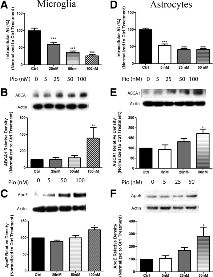
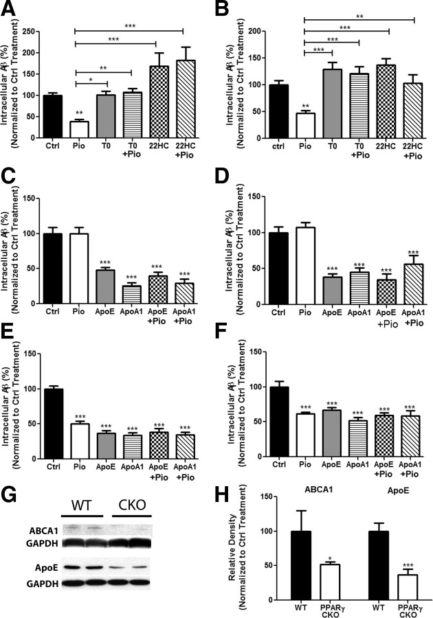
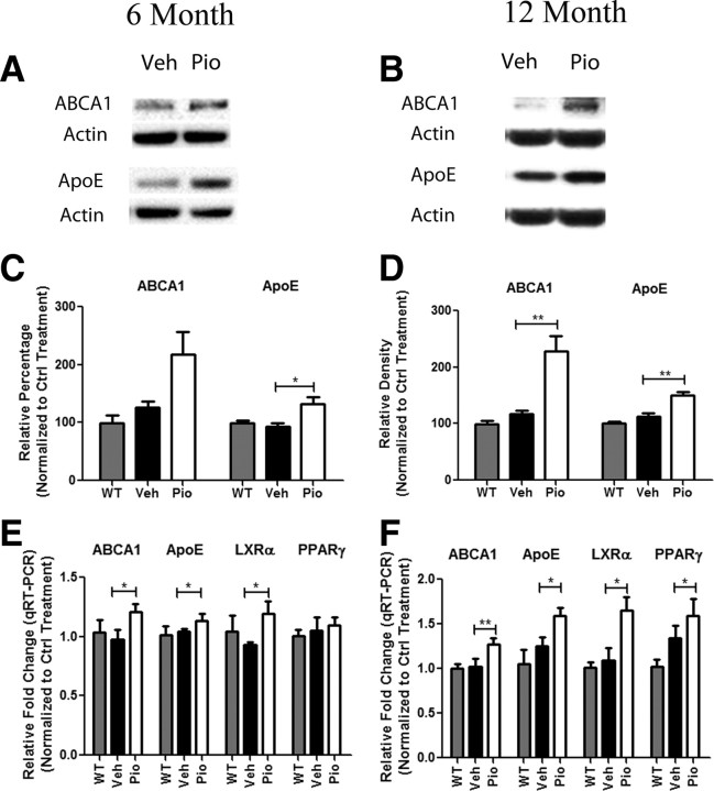
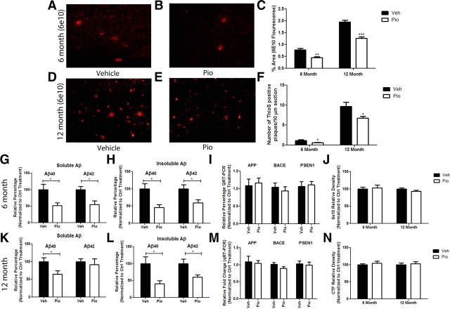
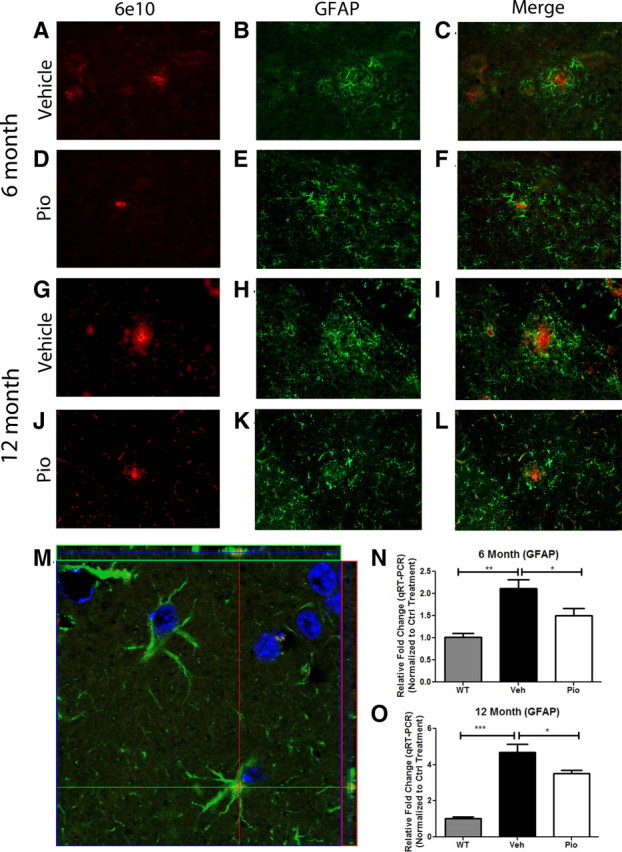
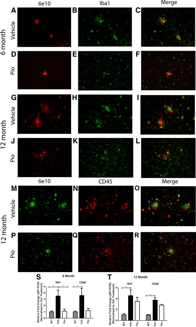
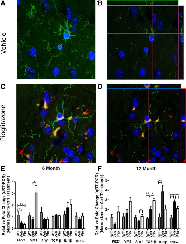
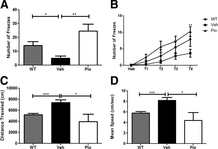
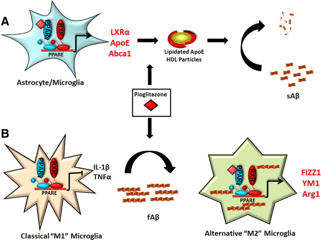
Alzheimer's disease is associated with a disruption of amyloid β (Aβ) homeostasis, resulting in the accumulation and subsequent deposition of Aβ peptides within the brain. The peroxisome proliferator-activated receptor-γ (PPARγ) is a ligand-activated nuclear receptor that acts in a coupled metabolic cycle with Liver X Receptors (LXRs) to increase brain apolipoprotein E (apoE) levels. apoE functions to promote the proteolytic clearance of soluble forms of Aβ, and we found that the synthetic PPARγ agonist, pioglitazone, stimulated Aβ degradation by both microglia and astrocytes in an LXR and apoE-dependent manner. Remarkably, a brief 9 d oral treatment of APPswe/PS1Δe9 mice with pioglitazone resulted in dramatic reductions in brain levels of soluble and insoluble Aβ levels which correlated with the loss of both diffuse and dense-core plaques within the cortex. The removal of preexisting amyloid deposits was associated with the appearance of abundant Aβ-laden microglia and astrocytes. Pioglitazone treatment resulted in the phenotypic polarization of microglial cells from a proinflammatory M1 state, into an anti-inflammatory M2 state that was associated with enhanced phagocytosis of deposited forms of amyloid. The reduction in amyloid levels was associated with a reversal of contextual memory deficits in the drug-treated mice. These data provide a mechanistic explanation for how PPARγ activation facilitates amyloid clearance and supports the therapeutic utility of PPARγ agonists for the treatment of Alzheimer's disease.


Figures

Figure 1.
Figure 1.
Activation of PPARγ facilitates the expression of LXR target genes and promotes the degradation of Aβ. Primary microglia (A) or astrocytes (D) were isolated from wild-type mice and pretreated with either DMSO (Ctrl) or increasing concentrations of pioglitazone (Pio) for 24 h. The cells were then incubated with 2 μg/ml Aβ in the presence of DMSO or the indicated dose of pioglitazone for 24 h. Intracellular Aβ levels were quantified using ELISA for Aβ42 and normalized to total protein (mean ±SEM, ***p < 0.001). Primary microglia (B, C) or astrocytes (E, F) were treated with DMSO or increasing doses of Pio for 24 h. Cellular lysates were subjected to SDS-PAGE, and Western analysis for abca1, apoe, or actin was performed (mean ± SEM, Student's t test, *p < 0.05, **p < 0.01). Quantification of three separate experiments has been graphed and representative Western blots have been included with each dataset. These data are represented as a percentage of DMSO-treated control samples.
Figure 2.
Figure 2.
PPARγ-mediated degradation of Aβ is dependent on induction of ApoE through the stimulation of LXR and PPAR pathways. Primary wild-type microglia (A) or astrocytes (B) were treated for 24 h with pioglitazone or DMSO followed by the addition of soluble Aβ42 (2 μg/ml) for 24 h. The cells were pretreated with antagonists for PPARγ (T0070907, 10 nm) or LXR (22S hydroxycholesterol, 10 μm) for 2 h. Intracellular Aβ was measured by ELISA. apoe knock-out microglia (C) or astrocytes (D), or wild-type microglia (E) or astrocytes (F) were pretreated for 24 h with DMSO or drug, followed by the addition of soluble Aβ42 and exogenously supplied apoE (1 μg/ml) or apoA1 (2 μg/ml). Remaining intracellular Aβ was measured using ELISA. These data are represented as a percentage of DMSO-treated control samples (mean ± SEM, Student's t test, *p < 0.05, **p < 0.01, ***p < 0.001, n ≥ 3). Cortical homogenates obtained from PPARγ conditional knock-out animals were analyzed by Western blot analysis for ABCA1, apoE, and actin (G) and quantified (H) (mean ± SEM, Student's t test, *p < 0.05, ***p < 0.001, n ≤ 6 animals per genotype).
Figure 3.
Figure 3.
PPARγ activation drives the expression of LXR target genes in vivo. Six (A, C)- and 12 (B, D)-month-old APP/PS1 mice were gavaged orally with 80 mg · kg−1 · d−1 pioglitazone (Pio) or vehicle (H2O) for 9 d. Cortical homogenates were then analyzed by Western blot for levels of abca1, apoe, and actin (A, B) and quantified (C, D). (mean ± SEM, *p < 0.05, **p < 0.01, n ≥ 6 animals/treatment). Quantitative PCR results for LXR target genes from cortices of 6 (E)- or 12 (F)-month-old APP/PS1 animals treated with pioglitazone (Pio) or vehicle (Veh) or wild-type animals treated with vehicle (WT) for 9 d (mean ± SEM, Student's t test, *p < 0.05, **p < 0.01, n ≤ 13 animals/group). Fold change is reported as a percentage of vehicle-treated WT animals.
Figure 4.
Figure 4.
Pioglitazone induces the clearance of amyloid in APP/PS1 mice. APP/PS1 mice were gavaged for 9 d with pioglitazone (80 mg · kg−1 · d−1) or vehicle (water). Plaque pathology was evaluated in the cortex of vehicle- or pioglitazone-treated 6 month (A–C, F–H) or 12 month (C–F, K, L) animals by staining with 6E10 (anti-Aβ antibody) (A, B, D, E) or thioflavin S. Quantification of 6E10+ plaque area (C) and number of thioS-positive plaques (F). Soluble and insoluble levels of Aβ40 and Aβ42 were measured by ELISA in 6 (G, H) and 12 (K, L) month animals. Real-time quantification of mRNA levels of app and the APP proteases bace and psen1 were measured in 6 (I) and 12 (M) month animals to determine whether pioglitazone treatment altered the expression of enzymes involved in APP processing. Cortical homogenates were evaluated for protein levels of APP and APP C-terminal fragments to determine whether PPARγ activation affected APP levels or processing in 6 month (J) and 12 month (N) animals (mean ± SEM, Student's t test, *p < 0.05, **p < 0.01, ***p < 0.001, n ≥ 6 animals/group).
Figure 5.
Figure 5.
PPARγ suppresses astrocytosis in APP/PS1 animals. GFAP and 6E10 immunochemistry on coronal sections from 6 (A–F)- or 12 (G–L)- month-old APP/PS1 animals treated with pioglitazone (D–F, J–L) or vehicle (A–C, G–I) for 9 d. Magnification 20×. Representative image of astrocytes from the cortex of 12 month pioglitazone-treated animal (M) (red, 6E10; green, GFAP; blue, DAPI; 100× magnification). Quantitative real-time PCR in vehicle-treated WT or APP/PS1 animals or pioglitazone-treated APP/PS1 animals for GFAP in 6 (N)- or 12 (O)-month-old animals (mean ± SEM, Student's t test, *p < 0.05, **p < 0.01, ***p < 0.001, n = 8–13 animals/group).
Figure 6.
Figure 6.
Pioglitazone suppresses microglial activation surrounding amyloid plaques in APP/PS1 animals. Cortical microglia were detected by Iba1 staining and Aβ by 6E10 immunochemistry on coronal sections from 6 (A–F)- or 12 (G–L)-month-old vehicle (A–C, G–I)- or pioglitazone (D–F, J–L)-treated APP/PS1 animals. Cortical CD45 and 6E10 staining in 12-month-old vehicle (M–O)- or pioglitazone (P–R)-treated animals. Real-time PCR quantification of transcript levels of Iba1 or CD45 in 6 (S)- and 12 (T)-month-old pioglitazone or vehicle-treated APP/PS1 animals (mean ± SEM, Student's t test, *p < 0.05, n ≤ 13 animals/group).
Figure 7.
Figure 7.
Pioglitazone treatment enhances microglial phagocytosis of Aβ. Representative images from vehicle (A, B)- or pioglitazone (C, D)-treated 12-month-old APP/PS1 animals. A and C represent projection images of z stacks (B, D). Green, Iba1; red, 6E10; blue, DAPI. Images were acquired using a Zeiss LSM 510 confocal microscope, 100× magnification. Six (E)- and 12 (F)-month-old APP/PS1 mice were gavaged orally with 80 mg · kg−1 · d−1 pioglitazone (Pio) or vehicle (Veh) for 9 d and WT animals were gavaged with H2O (WT). Quantitative real-time PCR was performed to assess transcript levels of M1 (classical; IL-1β, TNFα) or M2 (alternative; FIZZ1, YM1, Arg1, TGF-β) activation markers.
Figure 8.
Figure 8.
PPARγ activation reverses cognitive deficits in 12-month-old APP/PS1 animals. Contextual fear conditioning was examined in 12-month-old vehicle- or pioglitazone (80 mg · kg−1 · d−1)-treated APP/PS1 animals (A) (mean ± SEM, Student's t test, *p < 0.05, **p < 0.01, n = 7–10 animals/group). Number of freezes is shown as a function of training periods for contextual fear conditioning (two-way ANOVA, Bonferroni post hoc test, **p < 0.01) B–D, Measurement of distance traveled (C) and mean speed (D) in open-field analysis of vehicle-treated WT and APP/PS1- and pioglitazone-treated APP/PS1 animals.
Figure 9.
Figure 9.
PPARγ activation promotes amyloid clearance from the AD brain. We have shown that PPARγ activation, using the synthetic agonist pioglitazone, promotes the clearance of both soluble and fibrillar amyloid species from the AD brain via distinct mechanisms. PPARγ promotes the degradation of sAβ species through the induction of LXR target genes apoE and abca1. This increases the pool of lipidated apoE particles which then facilitate the proteolytic clearance of sAβ as we have previously described (A; Jiang et al., 2008). Additionally, PPARγ activation induces the phenotypic conversion of the classically activated microglia to an alternative activation state. This conversion suppresses the production of inflammatory cytokines such as IL-1β and TNFα and promotes the expression of genes involved in tissue repair and phagocytosis (FIZZ1, YM1, Arg1, TGF-β), allowing microglial cells to regain their capacity to take up and degrade fibrillar Aβ species (B).

References

    1. Akiyama H, Arai T, Kondo H, Tanno E, Haga C, Ikeda K. Cell mediators of inflammation in the Alzheimer disease brain. Alzheimer Dis Assoc Disord. 2000a;14(Suppl 1):S47–S53. - PubMed
    1. Akiyama H, Barger S, Barnum S, Bradt B, Bauer J, Cole GM, Cooper NR, Eikelenboom P, Emmerling M, Fiebich BL, Finch CE, Frautschy S, Griffin WS, Hampel H, Hull M, Landreth G, Lue L, Mrak R, Mackenzie IR, McGeer PL, et al. Inflammation and Alzheimer's disease. Neurobiol Aging. 2000b;21:383–421. - PMC - PubMed
    1. al-Ali SY, al-Hussain SM. An ultrastructural study of the phagocytic activity of astrocytes in adult rat brain. J Anat. 1996;188:257–262. - PMC - PubMed
    1. Beaven SW, Tontonoz P. Nuclear receptors in lipid metabolism: targeting the heart of dyslipidemia. Annu Rev Med. 2006;57:313–329. - PubMed
    1. Bolmont T, Haiss F, Eicke D, Radde R, Mathis CA, Klunk WE, Kohsaka S, Jucker M, Calhoun ME. Dynamics of the microglial/amyloid interaction indicate a role in plaque maintenance. J Neurosci. 2008;28:4283–4292. - PMC - PubMed

Publication types

MeSH terms