. 2012 Sep;18(9):1702-15.
doi: 10.1261/rna.034314.112. Epub 2012 Jul 26.

Multiple binding of repressed mRNAs by the P-body protein Rck/p54

Affiliations

Multiple binding of repressed mRNAs by the P-body protein Rck/p54

Michèle Ernoult-Lange et al. RNA. 2012 Sep.

Abstract

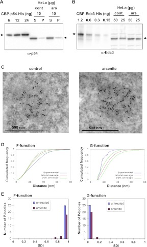
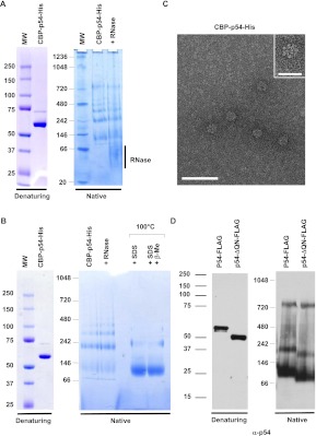
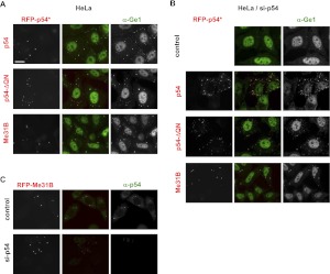
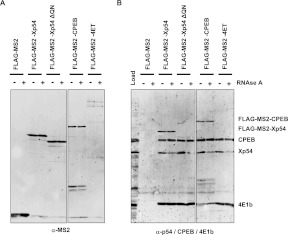
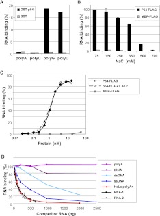
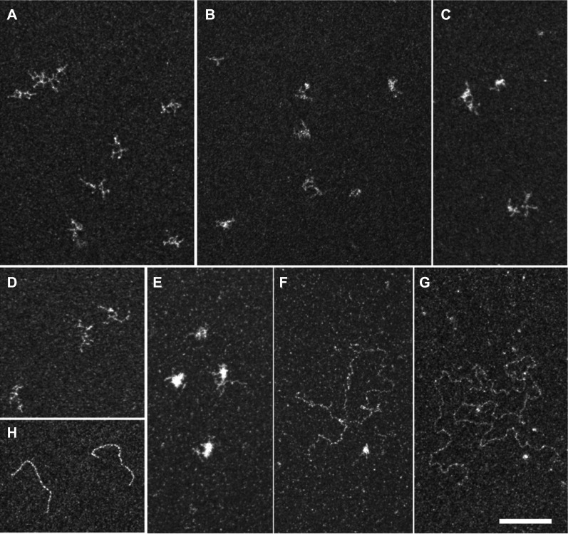
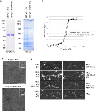
Translational repression is achieved by protein complexes that typically bind 3' UTR mRNA motifs and interfere with the formation of the cap-dependent initiation complex, resulting in mRNPs with a closed-loop conformation. We demonstrate here that the human DEAD-box protein Rck/p54, which is a component of such complexes and central to P-body assembly, is in considerable molecular excess with respect to cellular mRNAs and enriched to a concentration of 0.5 mM in P-bodies, where it is organized in clusters. Accordingly, multiple binding of p54 proteins along mRNA molecules was detected in vivo. Consistently, the purified protein bound RNA with no sequence specificity and high nanomolar affinity. Moreover, bound RNA molecules had a relaxed conformation. While RNA binding was ATP independent, relaxing of bound RNA was dependent on ATP, though not on its hydrolysis. We propose that Rck/p54 recruitment by sequence-specific translational repressors leads to further binding of Rck/p54 along mRNA molecules, resulting in their masking, unwinding, and ultimately recruitment to P-bodies. Rck/p54 proteins located at the 5' extremity of mRNA can then recruit the decapping complex, thus coupling translational repression and mRNA degradation.


Figures

FIGURE 1.
FIGURE 1.
Quantitation of the Rck/p54 protein in the cytoplasm and in P-bodies. HeLa cells were treated or not with arsenite to increase the number of P-bodies. (A) Quantitation of Rck/p54 in HeLa cells. Fifteen micrograms of soluble (S) and insoluble (P) proteins were analyzed along with 6–24 ng of recombinant CBP-p54-His protein by Western blotting with an anti-p54 antibody. The arrows indicate the position of the recombinant and human Rck/p54 proteins. (B) Quantitation of Edc3 in HeLa cells. A total of 50 and 25 μg of proteins were analyzed along with 0.15–1.2 ng of recombinant CBP-Edc3-His protein by Western blotting with an anti-Edc3 antibody. The arrows indicate the position of the recombinant and human Edc3 proteins. (C) Concentration of Rck/p54 in P-bodies. HeLa cells fixed with paraformaldehyde were embedded in Lowicryl and immunostained with anti-p54 antibodies coupled to 10-nm gold particles. The labeling is concentrated over the electron-dense P-body. (D) Statistical analysis of Rck/p54 spatial distribution in P-bodies. Immunoelectron microscopy images were analyzed with distance functions F (left) and G (right). The curves obtained for the P-body (C, left) (in blue) were compared with the average curve (in red) and its 95% confidence envelope (in green) obtained with random distributions of the same number of gold-particles in the same contour (hard-core binomial model). (E) Population distribution of SDIs calculated for functions F (left) and G (right), for 46 P-bodies from untreated and arsenite-treated cells, as indicated.
FIGURE 2.
FIGURE 2.
RNA-independent oligomerization of recombinant Rck/p54 in vitro. (A) Oligomerization of purified CBP-p54-His. A total of 2 μg of purified recombinant CBP-p54-His was separated on denaturing (left) and native (right) gels, and stained by Coomassie. Where indicated, the protein was treated with RNase A prior to migration. (B) Aberrant migration of Rck/p54 in native gels. A total of 2 μg of purified recombinant CBP-p54-His was separated on denaturing (left) and native (right) gels, stained with Coomassie. Where indicated, the protein was treated with RNase prior to purification, or heated in the presence of SDS with or without β-mercaptoethanol prior to migration. (C) Visualization of the purified protein in electron microscopy. The protein preparation analyzed in A was spread on grids and stained with uranyl acetate prior to observation by electron microscopy in filtered zero loss mode. (Main panel) Magnification ×151,000; bar, 50 nm. (Inset) Magnification ×241,500; bar, 20 nm. (D) Oligomerization is independent of the QN-rich extension of the protein. A total of 50 ng of purified recombinant FLAG-tagged wild-type (p54-FLAG) and truncated (p54-ΔQN-FLAG) Rck/p54 proteins were separated on denaturing (left) and native (right) gels, and analyzed by Western blotting with anti-p54 antibodies.
FIGURE 3.
FIGURE 3.
The Rck/p54 QN-rich extension is dispensable for P-body assembly. (A) Localization in pre-existing P-bodies in the absence of the QN-rich region. HeLa cells were transfected with expression vectors encoding RFP-tagged human wild-type (p54) and truncated (p54-ΔQN) Rck/p54, and Drosophila Me31B. After 40 h, P-bodies were stained with anti-Ge1 antibodies and observed by fluorescence microscopy. Bar, 20 μm. (B,C) De novo assembly of P-bodies in the absence of the QN-rich region. HeLa cells were transfected with si-p54 to deplete Rck/p54 and suppress P-bodies. Twenty-four hours later, cells were transfected as described in A and stained with anti-Ge1 antibodies. (C) P-bodies assembled with Me31B do not contain human Rck/p54. P-bodies were assembled with Me31B as described in B and stained with anti-p54 antibodies.
FIGURE 4.
FIGURE 4.
Multiple Rck/p54 binding along mRNA molecules. Xenopus oocytes were injected with an mRNA encoding the indicated proteins. Sixteen hours later, cells were lysed and treated or not with RNAse before immunoprecipitation with anti-FLAG antibody. Immunoprecipitates were analyzed by Western blotting with anti-MS2 (A) or a combination of anti-CPEB, anti-p54, and anti-eIF4E1b antibodies (B). Note that the MS2-Xp54-ΔQN is not detected by the anti-p54 antibody, which recognizes an epitope in the QN region.
FIGURE 5.
FIGURE 5.
High-affinity and sequence-independent binding of Rck/p54 to RNA. (A) Rck/p54 binding to RNA homopolymers. GST-tagged Rck/p54 and control GST proteins bound to glutathione beads were incubated with radiolabeled RNA homopolymers. The histogram represents the percentage of RNA retained on the beads with the proteins. (B) Sensitivity of RNA binding to salt concentration. Radiolabeled RNA (0.25 ng) was incubated with purified FLAG-tagged Rck/p54 and control MBP proteins (120 ng) in the presence of indicated salt concentrations. RNA binding was assessed by filter-retention assay. The histogram represents the percentage of RNA retained on the filter with the proteins. (C) ATP-independent and high-affinity RNA binding. Radiolabeled RNA (0.25 ng) was incubated with increasing concentrations of the purified FLAG-tagged Rck/p54 and MBP proteins, in the presence or absence of ATP. RNA binding was assessed as in B. (D) Preferential and sequence-independent RNA binding. Radiolabeled RNA (0.25 ng) was incubated with the purified FLAG-tagged Rck/p54 (120 ng) in the presence of various amounts of the indicated RNA and DNA competitors. RNA binding was assessed as in B.
FIGURE 6.
FIGURE 6.
Electron microscopy imaging of RNA bound to Rck/p54. RNA was incubated alone (A,D) or with a 40 molar excess of the double-purified CBP-p54-His (B,C,E–G), in the absence (A–C) or presence (D–G) of ATP. The mixture was spread on grids and stained with uranyl acetate prior to observation by electron microscopy in filtered crystallographic dark field mode. RNA molecules alone were highly branched, independently of the presence of ATP (A,D). Upon protein binding in the absence of ATP, only branched RNA molecules were observed, as seen in two representative fields (B,C). In the presence of ATP, a mixture of ∼80% branched (E) and 20% relaxed (F,G) RNA molecules were observed. The characteristic spreading of double-stranded DNA in the same condition is shown for comparison (H). Magnification ×69,100; bar, 100 nm.
FIGURE 7.
FIGURE 7.
RNA relaxing in the presence of Rck/p54 is independent of ATP hydrolysis. (A) Oligomerization of Rck/p54 DQAD mutant. A total of 2 μg of purified recombinant CBP-p54-His proteins, with or without an E/Q mutation in the DEAD motif, were separated on denaturing (left) and native (right) gels as in Figure 2A. (B) Visualization of Rck/p54 DQAD protein in electron microscopy. Both wild-type and mutant CBP-His-tagged proteins were visualized in electron microscopy as in Figure 2C. (Main panels) Magnification ×69,100; bar, 100 nm. (Insets) Magnification ×151,000; bar, 20 nm. (C) RNA-binding properties of Rck/p54 DQAD mutant. RNA binding was measured in the presence and absence of ATP using FLAG-tagged protein and filter retention assay, as in Figure 5C. (D) Electron microscopy imaging of RNA bound to Rck/p54 in the presence of AMP-PNP, or to Rck/p54 DQAD mutant. RNA was incubated with a 40 molar excess of the double-purified wild-type or mutant CBP-His-tagged proteins in the absence or presence of ATP, AMP-PNP, or ADP, as indicated. The mixture was spread and observed as in Figure 6. RNA alone is also shown. Magnification ×69,100; bar, 100 nm.
FIGURE 8.
FIGURE 8.
Proposed model for the participation of Rck/p54 to translational repression and mRNA decay. Translational repression is initiated by the specific binding of protein X and its partners, including Rck/p54 (the exact stoichiometry of the repressor complex is not represented here). This triggers the sequence-independent binding of Rck/p54 along the mRNA. RNA relaxing is then favored by the presence of ATP. These extended mRNP complexes are then recruited to P-bodies, giving rise to the Rck/p54-rich fibers observed in electron microscopy. They can either return to translation if Rck/p54 is released, or the Rck/p54 located close to the 5′ cap can recruit the decapping complex, leading to mRNA degradation.

References

    1. Andrey P, Kiêu K, Kress C, Lehmann G, Tirichine L, Liu Z, Biot E, Adenot PG, Hue-Beauvais C, Houba-Herin N, et al. 2010. Statistical analysis of 3D images detects regular spatial distributions of centromeres and chromocenters in animal and plant nuclei. PLoS Comput Biol 6: e1000853 doi: 10.1371/journal.pcbi.1000853 - PMC - PubMed
    1. Ballut L, Marchadier B, Baguet A, Tomasetto C, Seraphin B, Le Hir H 2005. The exon junction core complex is locked onto RNA by inhibition of eIF4AIII ATPase activity. Nat Struct Mol Biol 12: 861–869 - PubMed
    1. Boag PR, Atalay A, Robida S, Reinke V, Blackwell TK 2008. Protection of specific maternal messenger RNAs by the P body protein CGH-1 (Dhh1/RCK) during Caenorhabditis elegans oogenesis. J Cell Biol 182: 543–557 - PMC - PubMed
    1. Boulton AA, Baker GB, Campagnoni AT, ed. 1990. Molecular neurobiological techniques. In Neuromethods, Vol 16. Humana Press, Clifton, NJ
    1. Chu CY, Rana TM 2006. Translation repression in human cells by microRNA-induced gene silencing requires RCK/p54. PLoS Biol 4: e210 doi: 10.1371/journal.pbio.0040210 - PMC - PubMed

Publication types

LinkOut - more resources