. 2013 Jan;31(1):91-8.
doi: 10.1002/jor.22191. Epub 2012 Jul 26.

Substance P enhances collagen remodeling and MMP-3 expression by human tenocytes

Affiliations

Substance P enhances collagen remodeling and MMP-3 expression by human tenocytes

Gloria Fong et al. J Orthop Res. 2013 Jan.

Abstract

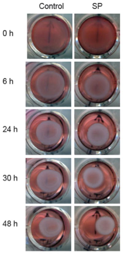
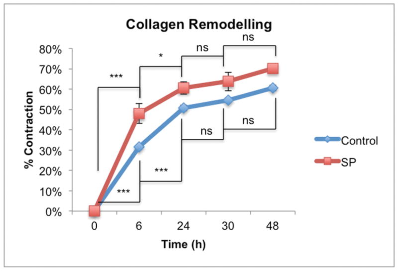
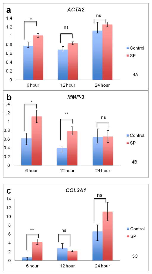
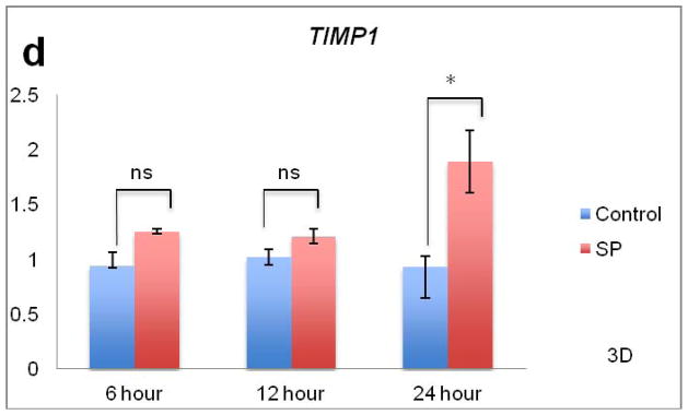
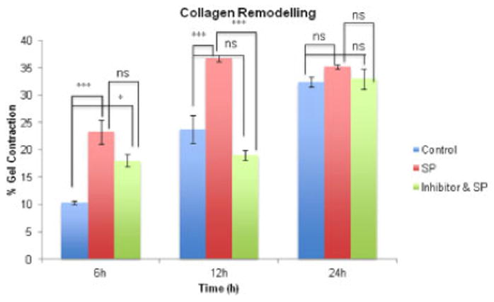
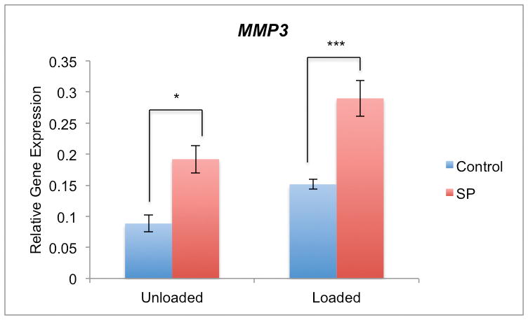
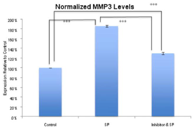
The loss of collagen organization is considered a hallmark histopathologic feature of tendinosis. At the cellular level, tenocytes have been shown to produce signal substances that were once thought to be restricted to neurons. One of the main neuropeptides implicated in tendinosis, substance P (SP), is known to influence collagen organization, particularly after injury. The aim of this study was to examine the influence of SP on collagen remodeling by primary human tendon cells cultured in vitro in three-dimensional collagen lattices. We found that SP stimulation led to an increased rate of collagen remodeling mediated via the neurokinin-1 receptor (NK-1 R), the preferred cell receptor for SP. Gene expression analysis showed that SP stimulation resulted in significant increases in MMP3, COL3A1 and ACTA2 mRNA levels in the collagen lattices. Furthermore, cyclic tensile loading of tendon cell cultures along with the administration of exogenous SP had an additive effect on MMP3 expression. Immunoblotting confirmed that SP increased MMP3 protein levels via the NK-1 R. This study indicates that SP, mediated via NK-1 R, increases collagen remodeling and leads to increased MMP3 mRNA and protein expression that is further enhanced by cyclic mechanical loading.


Figures

Figure 1
Figure 1
Remodeling of control and SP treated (10−7 M) tenocytes.
Figure 2
Figure 2
Remodeling by control and SP treated (10−7 M) tenocytes increased in a time-dependent manner (p<0.001, two-way ANOVA). The rate of remodeling by SP treated tenocytes was greater than the control group (p<0.01, two-way ANOVA). For the control and SP treated groups most of the remodeling occurred within 24 hours post-release (0–6h*** p<0.001 for both control and SP treated groups and 6–24h ***p<0.001 and *p<0.05 for control and SP treated respectively, two-way ANOVA, followed by Bonferroni post-hoc test) after which there was no difference between the two groups at the 30 and 48-hour time points.
Figure 3
Figure 3
Effects of SP on collagen remodeling and mRNA levels. Figure 3A. For ACTA2, expression increased in both control and SP treated tenocytes (p<0.01, two-way ANOVA) while SP further increased ACTA2 expression compared to control (p<0.05, two-way ANOVA). There was an interaction between time and treatment. At the 6 hour time point, SP significantly upregulated ACTA2 compared to control (*p<0.05, independent samples t-test). Figure 3B. There was an increase in MMP3 expression due to SP treatment (p<0.001, two-way ANOVA). There was no interaction between time and treatment. SP led to significant upregulation of MMP3 at 6 and 12 hours compared to the control (*p<0.05 and **p<0.01, independent samples t-test respectively). Figure 3C. For COL3A1, expression increased in both control and SP treated tenocytes (p<0.05, two-way ANOVA). There was an interaction between time and treatment. At the 6 hour time point, SP significantly upregulated COL3A1 compared to the control (**p<0.01, independent samples t-test). Figure 3D. There was an increase in TIMP1 expression due to SP treatment (p<0.001, two-way ANOVA). There was an interaction between time and treatment. SP led to significant upregulation of TIMP1 at the 24 hour time point (*p<0.05, independent samples t-test).
Figure 3
Figure 3
Effects of SP on collagen remodeling and mRNA levels. Figure 3A. For ACTA2, expression increased in both control and SP treated tenocytes (p<0.01, two-way ANOVA) while SP further increased ACTA2 expression compared to control (p<0.05, two-way ANOVA). There was an interaction between time and treatment. At the 6 hour time point, SP significantly upregulated ACTA2 compared to control (*p<0.05, independent samples t-test). Figure 3B. There was an increase in MMP3 expression due to SP treatment (p<0.001, two-way ANOVA). There was no interaction between time and treatment. SP led to significant upregulation of MMP3 at 6 and 12 hours compared to the control (*p<0.05 and **p<0.01, independent samples t-test respectively). Figure 3C. For COL3A1, expression increased in both control and SP treated tenocytes (p<0.05, two-way ANOVA). There was an interaction between time and treatment. At the 6 hour time point, SP significantly upregulated COL3A1 compared to the control (**p<0.01, independent samples t-test). Figure 3D. There was an increase in TIMP1 expression due to SP treatment (p<0.001, two-way ANOVA). There was an interaction between time and treatment. SP led to significant upregulation of TIMP1 at the 24 hour time point (*p<0.05, independent samples t-test).
Figure 4
Figure 4
Collagen remodeling 6, 12 and 24 hours after release of lattices (control-, SP- (10−7 M), and NK-1 R inhibitor (10−6 M) & SP- (10−7 M)- treated). The extent of remodeling in the SP treated group was increased compared to control at 6 and 12 hours (6h ***p<0.001, 12h ***p<0.001, 2-way ANOVA, followed by Bonferroni post-hoc test). The inhibitor temporary blocked the effects of SP at 12h but the effect was incomplete at 6h compared to control (*p<0.05, 2-way ANOVA, followed by Bonferroni post-hoc test). At 12h, treatment with SP resulted in significant increase in remodeling while treatment of the inhibitor blocked the effect (***p<0.001, 2-way ANOVA, followed by Bonferroni post-hoc test).
Figure 5
Figure 5
qPCR of MMP3 mRNA in cultured human primary Achilles tendon cells. MMP3 expression is significantly different between unloaded and loaded tenocytes (p<0.01, two-way ANOVA). The mean level of MMP3 expression is elevated for both unloaded and loaded samples following SP (10−7 M) stimulation (p<0.001, two-way ANOVA). For both the unloaded and loaded groups, there was approximately a two-hold increase in MMP3 expression due to SP (*p<0.5 and ***p<0.001 respectively, two-way ANOVA, followed by Bonferroni post-hoc test).
Figure 6
Figure 6
Effect of SP on MMP3 protein level. Analysis of MMP3 after 12 hours of incubation with SP (10−7 M), without SP (control), and with SP and the NK-1 R blocker (10−6 M), as measured by normalized band intensity. MMP3 was significantly increased after incubation with SP (***p<0.001, one-way ANOVA, followed by Bonferroni post-hoc test) and this increase was much less obvious in the presence of the NK-1 R antagonist (***p<0.001, one-way ANOVA, followed by Bonferroni post-hoc test) when both are compared to the control. As well, the NK-1 R antagonist resulted in significant decreased in MMP3 protein level relative to treatment with SP (***p<0.001, one-way ANOVA, followed by Bonferroni post-hoc test).

References

    1. Khan KM, Cook JL, Bonar F, et al. Histopathology of common tendinopathies. Update and implications for clinical management. Sports Med. 1999;27:393–408. - PubMed
    1. Sivaguru M, Durgam S, Ambekar R, et al. Quantitative analysis of collagen fiber organization in injured tendons using Fourier transform-second harmonic generation imaging. Opt Express. 2010;24:24983–93. - PubMed
    1. Riley G. Chronic tendon pathology: molecular basis and therapeutic implications. Expert Reviews in Molecular Medicine. 2005;5:1–25. - PubMed
    1. Forsgren S, Alfredson H, Bjur D, et al. Novel information on the non-neuronal cholinergic system in orthopedics provides new possible treatment strategies for inflammatory and degenerative diseases. Orthop Rev (Pavia) 2009;1:e11. - PMC - PubMed
    1. Bjur D, Danielson P, Alfredson H, Forsgren S. Presence of a non-neuronal cholinergic system and occurrence of up- and down-regulation in expression of M2 muscarinic acetylcholine receptors: new aspects of importance regarding Achilles tendon tendinosis (tendinopathy) Cell Tissue Res. 2008;2:385–400. - PubMed

Publication types

MeSH terms