. 2012 Jul 26;15(8):1068-73.
doi: 10.1038/nn.3155.

P2X4R+ microglia drive neuropathic pain

Affiliations

P2X4R+ microglia drive neuropathic pain

Simon Beggs et al. Nat Neurosci. .

Abstract

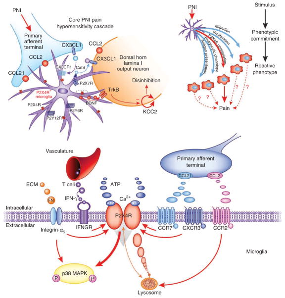
Neuropathic pain, the most debilitating of all clinical pain syndromes, may be a consequence of trauma, infection or pathology from diseases that affect peripheral nerves. Here we provide a framework for understanding the spinal mechanisms of neuropathic pain as distinct from those of acute pain or inflammatory pain. Recent work suggests that a specific microglia response phenotype characterized by de novo expression of the purinergic receptor P2X4 is critical for the pathogenesis of pain hypersensitivity caused by injury to peripheral nerves. Stimulating P2X4 receptors initiates a core pain signaling pathway mediated by release of brain-derived neurotrophic factor, which produces a disinhibitory increase in intracellular chloride in nociceptive (pain-transmitting) neurons in the spinal dorsal horn. The changes caused by signaling from P2X4R(+) microglia to nociceptive transmission neurons may account for the main symptoms of neuropathic pain in humans, and they point to specific interventions to alleviate this debilitating condition.


Conflict of interest statement

The authors declare no competing financial interests.

Figures

Figure 1
Figure 1
Microglia; microdomains and proliferation after nerve injury. (a) Transverse section of the lumbar spinal cord showing the characteristic proliferation of microglia (immunostained with iba1, green) around the central terminals of primary afferents whose peripheral axons have been severed (right side). Scale bar, 100 μm. Insets: confocal images showing the characteristic morphology in the absence of peripheral nerve injury (left) and the morphological change that occurs after injury (right). Scale bar, 10 μm. (b) In the normal brain and spinal cord, microglia have a grid-like distribution. Two-photon microscopy image (top) shows the distribution in vivo of microglia in the parenchyma of the CNS of a CX3CR1GFP mouse (in which the Cx3cr1 gene, expressed specifically in microglia, has been replaced with a gene encoding green fluorescent protein). Three microglia have been surface-rendered using Volocity software (PerkinElmer) to show the adjacent, non-overlapping microdomains of individual cells (bottom); center, overlay. Scale bar, 30 μm.
Figure 2
Figure 2
The P2X4R+ microglial phenotype mediates a core pain hypersensitivity cascade following peripheral nerve injury. Chronic pain due to peripheral nerve injury is caused by amplification of input from the damaged nerve (blue) by the nociceptive network in the spinal cord. Neuronglia interactions in the spinal cord contribute to the resulting enhanced output of this network. Peripheral axotomy or nerve damage (PNI) stimulates changes in spinal microglia (purple) such that they adopt a phenotype characterized by increased expression of P2X4Rs (top left). CCL2, chemokine (C-C motif) ligand 2; CX3CL1, chemokine (C-X3-C motif) ligand 1; P2X7R, P2X purinoceptor 7; P2Y12R, P2Y purinoceptor 12; P2Y6R, P2Y purinoceptor 6; CatS, cathepsin S. The dotted rectangle is expanded in the bottom panel. This is one of several reactive phenotypes that microglia can adopt under pathological conditions (top right). Whether other phenotypes contribute to the induction and maintenance of neuropathic pain is not fully understood. Peripheral nerve injury modulates P2X4R expression by the release of various factors from the extracellular matrix, infiltrating T cells and injured primary afferents (bottom). P2X4Rs are localized predominantly to lysosomes and are trafficked to the cell surface (orange arrow). P2X4R activation by ATP leads to calcium inflow, phosphorylation (P) of p38 MAPK, and p38-mediated synthesis and release of BDNF from the microglia. BDNF then binds neuronal TrkB receptors in the spinal nociceptive network and in turn causes a downregulation of the potassium–chloride cotransporter KCC2, disturbing the chloride homeostasis of the neuron. The outcome is a net disinhibition of the intrinsic inhibitory system in the dorsal horn. ECM, extracellular matrix; IFN, interferon; IFNGR, interferon-γ receptor; CXCR3, chemokine (C-X-C motif) receptor 3; CCR7, C-C chemokine receptor type 7. Red arrows indicate increased expression and activation.

References

    1. Hanisch UK, Kettenmann H. Microglia: active sensor and versatile effector cells in the normal and pathologic brain. Nat Neurosci. 2007;10:1387–1394. - PubMed
    1. Tsuda M, et al. P2X4 receptors induced in spinal microglia gate tactile allodynia after nerve injury. Nature. 2003;424:778–783. - PubMed
    1. Ulmann L, et al. Up-regulation of P2X4 receptors in spinal microglia after peripheral nerve injury mediates BDNF release and neuropathic pain. J Neurosci. 2008;28:11263–11268. - PMC - PubMed
    1. Trang T, Beggs S, Wan X, Salter MW. P2X4-receptor-mediated synthesis and release of brain-derived neurotrophic factor in microglia is dependent on calcium and p38-mitogen-activated protein kinase activation. J Neurosci. 2009;29:3518–3528. - PMC - PubMed
    1. Nissl F. Ueber einige Beziehungen zur Nervenzellenerkrankungen und gliosen Erscheinungen bei verschiedenen Psychosen. Arch Psychiatr. 1899;32:656–676.

Publication types