Review
. 2012 Jul 26;15(8):1096-101.
doi: 10.1038/nn.3161.

Immune surveillance in the central nervous system

Affiliations
Review

Immune surveillance in the central nervous system

Shalina S Ousman et al. Nat Neurosci. .

Erratum in

  • Nat Neurosci. 2014 Sep;17(9):1286

Abstract

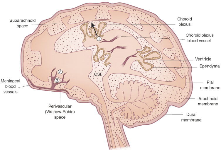
The CNS, which consists of the brain and spinal cord, is continuously monitored by resident microglia and blood-borne immune cells such as macrophages, dendritic cells and T cells to detect for damaging agents that would disrupt homeostasis and optimal functioning of these vital organs. Further, the CNS must balance between vigilantly detecting for potentially harmful factors and resolving any immunological responses that in themselves can create damage if left unabated. We discuss the physiological roles of the immune sentinels that patrol the CNS, the molecular markers that underlie their surveillance duties, and the consequences of interrupting their functions following injury and infection by viruses such as JC virus, human immunodeficiency virus, herpes simplex virus and West Nile virus.


Conflict of interest statement

The authors declare no competing financial interests.

Figures

Figure 1
Figure 1. Possible sites of immune cell entry into the CNS.
T cells and monocytes may gain access to the brain by crossing through the fenestrated blood vessels of the choroid plexus, across the ependymal layer and into the CSF (1); through the perivascular or Virchow-Robin space, where the meningeal blood vessels branch into the subarachnoid space (2); and directly into the CNS parenchyma through postcapillary venules (3).

References

    1. Carvalho-Tavares J, et al. A role for platelets and endothelial selectins in tumor necrosis factor-α–induced leukocyte recruitment in the brain microvasculature. Circ. Res. 2000;87:1141–1148. doi: 10.1161/01.RES.87.12.1141. - DOI - PubMed
    1. Hickey WF. Basic principles of immunological surveillance of the normal central nervous system. Glia. 2001;36:118–124. doi: 10.1002/glia.1101. - DOI - PubMed
    1. Prinz M, Priller J, Sisodia SS, Ransohoff RM. Heterogeneity of CNS myeloid cells and their roles in neurodegeneration. Nat. Neurosci. 2011;14:1227–1235. doi: 10.1038/nn.2923. - DOI - PubMed
    1. Ginhoux F, et al. Fate mapping analysis reveals that adult microglia derive from primitive macrophages. Science. 2010;330:841–845. doi: 10.1126/science.1194637. - DOI - PMC - PubMed
    1. Alliot F, Lecain E, Grima B, Pessac B. Microglial progenitors with a high proliferative potential in the embryonic and adult mouse brain. Proc. Natl. Acad. Sci. USA. 1991;88:1541–1545. doi: 10.1073/pnas.88.4.1541. - DOI - PMC - PubMed

Publication types

MeSH terms