. 2012 Aug 17;337(6096):843-6.
doi: 10.1126/science.1224352. Epub 2012 Jul 26.

Identification of the Cdc48•20S proteasome as an ancient AAA+ proteolytic machine

Affiliations

Identification of the Cdc48•20S proteasome as an ancient AAA+ proteolytic machine

Dominik Barthelme et al. Science. .

Abstract

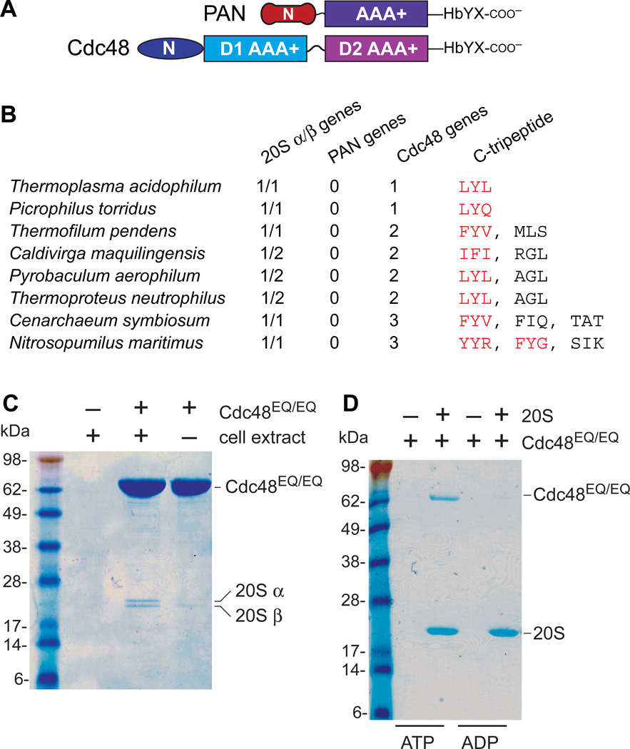
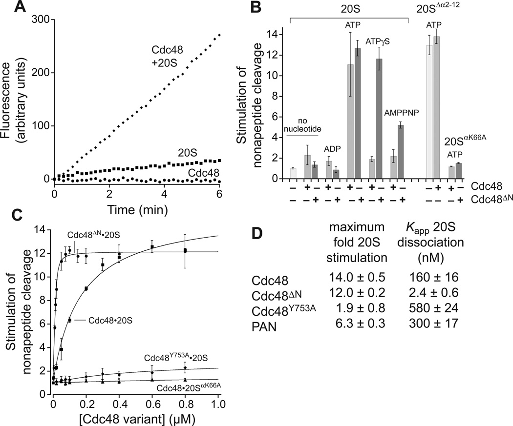
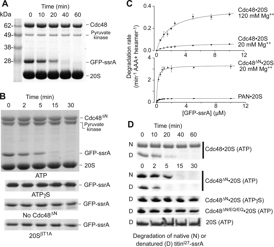
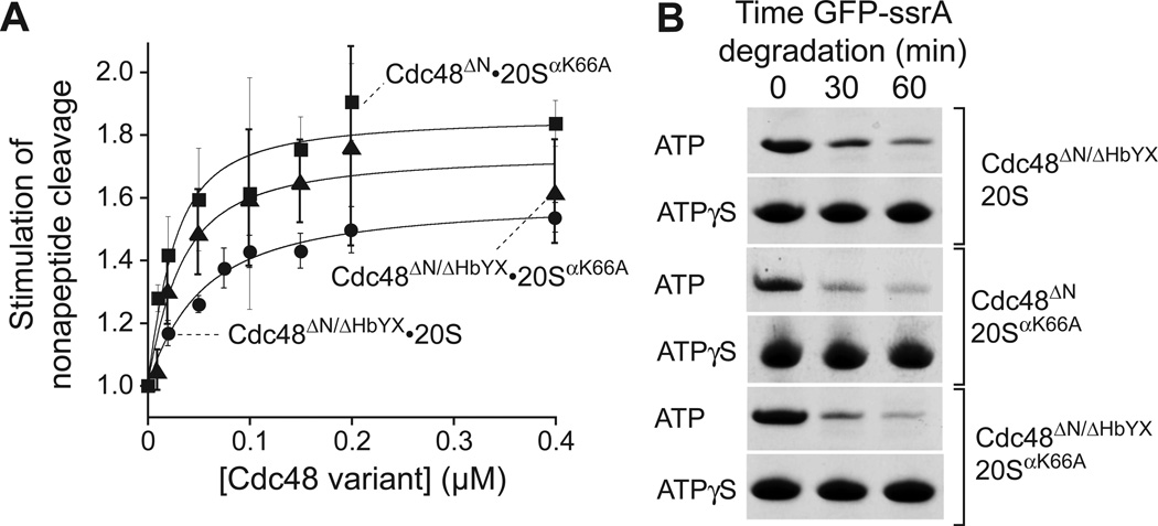
Proteasomes are the major energy-dependent proteolytic machines in the eukaryotic and archaeal domains of life. To execute protein degradation, the 20S core peptidase combines with the AAA+ ring of the 19S regulatory particle in eukarya or with the AAA+ proteasome-activating nucleotidase ring in some archaea. Here, we find that Cdc48 and 20S from the archaeon Thermoplasma acidophilum interact to form a functional proteasome. Cdc48 is an abundant and essential double-ring AAA+ molecular machine ubiquitously present in archaea, where its function has been uncertain, and in eukarya where Cdc48 participates by largely unknown mechanisms in diverse cellular processes, including multiple proteolytic pathways. Thus, proteolysis in collaboration with the 20S peptidase may represent an ancestral function of the Cdc48 family.


Figures

Fig. 1
Fig. 1. Cdc48 binds the 20S peptidase
(A) PAN and Cdc48 have distinct N domains but homologous AAA+ modules and HbYX motifs. (B) Examples of archaea in which PAN was absent but 20S and Ccd48 were present. Red Cdc48 tripeptides match HbYX or related sequences. (C) Sodium dodecyl sulfate polyacrylamide gel electrophoresis (SDS-PAGE) showed that purified T. acidophilum His6-Cdc48EQ/EQ pulled down two proteins from a T. acidophilum cell extract, which were identified as the 20S α and β subunits (Fig. S2). (D) Using purified T. acidophilum proteins, 20S with a His6-tagged β subunit pulled down roughly stoichiometric quantities of untagged Cdc48EQ/EQ in the presence of 2 mM ATP but not ADP.
Fig. 2
Fig. 2. Cdc48 activates 20S gate opening
(A) T. acidophilum Cdc48 (0.4 µM) and 20S (10 nM) cleaved a nonapeptide (10 µM) rapidly as assayed by increased fluorescence with 2 mM ATP. Cleavage by Cdc48 alone was undetected and by 20S alone was slow. (B) Cdc48 or Cdc48ΔN (0.4 µM) stimulation of nonapeptide cleavage by 20S, 20SΔα2–10, or 20SαK66A (10 nM) in the presence of different nucleotides (2 mM). Values are averages ± SD (N ≥ 3) divided by the 20S cleavage rate. (C) Nonapeptide cleavage by 20S or 20SαK66A (10 nM) as a function of increasing concentrations of Cdc48 or variants. Solid lines (R2 ≥ 0.98) are fits to a hyperbolic equation for all curves except Cdc48ΔN•20S, which was fit to a quadratic equation for near-stoichiometric binding. (D) Values for maximum stimulation of cleavage and Kapp were calculated from the fits in panel C or Fig. S4.
Fig. 3
Fig. 3. Protein degradation by the Cdc48•20S proteasome
(A) SDS-PAGE assay of GFP-ssrA (10 µM) degradation by Cdc48 (1 µM) and 20S (2 µM) with 2 mM ATP, 120 mM MgCl2, and an ATP-regeneration system. (B) Upper panel is SDS-PAGE assay of GFP-ssrA (5 µM) degradation by 20S (0.9 µM) and Cdc48ΔN (0.3 µM), 2 mM ATP, 20 mM MgCl2, and a regeneration system. Lower strips show GFP-ssrA in otherwise comparable assays using 2 mM ATPγS (no regeneration), without Cdc48ΔN, or with catalytically inactive 20SβT1A. (C) Michaelis-Menten plots of GFP-ssrA degradation by 20S (0.9 µM) and Cdc48 (0.3 µM; left panel) or 20S (0.9 µM) with Cdc48ΔN or PAN (0.3 µM; right panel). Values are averages ± SD (N ≥ 3). Fitted parameters are listed in Table S2. (D) Degradation of 10 µM native titinI27-ssrA or denatured CM-titinI27-ssrA. The top two gel strips show degradation by Cdc48 (1.2 µM) and 20S (0.4 µM) in 120 mM Mg++ buffer. The next four strips show degradation by Cdc48ΔN or Cdc48ΔN/EQ/EQ (1.2 µM) and 20S (0.4 µM) in 20 mM Mg++ buffer. The bottom strip shows degradation by 20S (0.4 µM) in 20 mM Mg++ buffer. Reactions contained 5 mM ATP and a regeneration system or 5 mM ATPγS without regeneration.
Fig. 4
Fig. 4. The HbYX motif is not required for 20S binding and degradation
(A) Stimulation of peptide cleavage by 20S or 20SαK66A (10 nM) as a function of Cdc48ΔN/HbYX or Cdc48ΔN concentration. Values are averages (N=3) ± SD. Solid lines (R2 ≥ 0.948) are fits to a quadratic equation for near-stoichiometric binding, yielding Kapp values of 41 ± 4 nM for Cdc48ΔN/HbYX•20S, 21 ± 9 nM for Cdc48ΔN•20SαK66A, and 15 ± 6 nM for Cdc48ΔN/HbYX•20SαK66A. (B) Gel strips showing ATP-dependent degradation of GFP-ssrA (5 µM) by Cdc48ΔN/ΔHbYX•20S, Cdc48ΔN•20SαK66A, or Cdc48ΔN/HbYX•20SαK66A. Assays were performed in 20 mM Mg++ using 5 mM ATP and regeneration or 5 mM ATPγS; 20S/20SαK66A (0.4 µM); Cdc48ΔN/Cdc48ΔN/HbYX (1.2 µM).

Comment in

References

    1. Sauer RT, Baker TA. AAA+ proteases: ATP-fueled machines of protein destruction. Annu. Rev. Biochem. 2011;80:587–612. - PubMed
    1. Voges D, Zwickl P, Baumeister W. The 26S proteasome: a molecular machine designed for controlled proteolysis. Annu. Rev. Biochem. 1999;68:1015–1068. - PubMed
    1. Lander GC, et al. Complete subunit architecture of the proteasome regulatory particle. Nature. 2012;482:186–191. - PMC - PubMed
    1. Zwickl P, Ng D, Woo KM, Klenk HP, Goldberg AL. An archaebacterial ATPase, homologous to ATPases in the eukaryotic 26 S proteasome, activates protein breakdown by 20 S proteasomes. J. Biol. Chem. 1999;274:26008–26014. - PubMed
    1. Zhou G, Kowalczyk D, Humbard MA, Rohatgi S, Maupin-Furlow JA. Proteasomal components required for cell growth and stress responses in the haloarchaeon Haloferax volcanii. J. Bacteriol. 2008;190:8096–8105. - PMC - PubMed

Publication types

MeSH terms