. 2012 Sep 1;189(5):2530-6.
doi: 10.4049/jimmunol.1200994. Epub 2012 Jul 25.

CD4+ T cell-dependent IFN-γ production by CD8+ effector T cells in Mycobacterium tuberculosis infection

Affiliations

CD4+ T cell-dependent IFN-γ production by CD8+ effector T cells in Mycobacterium tuberculosis infection

Tyler D Bold et al. J Immunol. .

Abstract

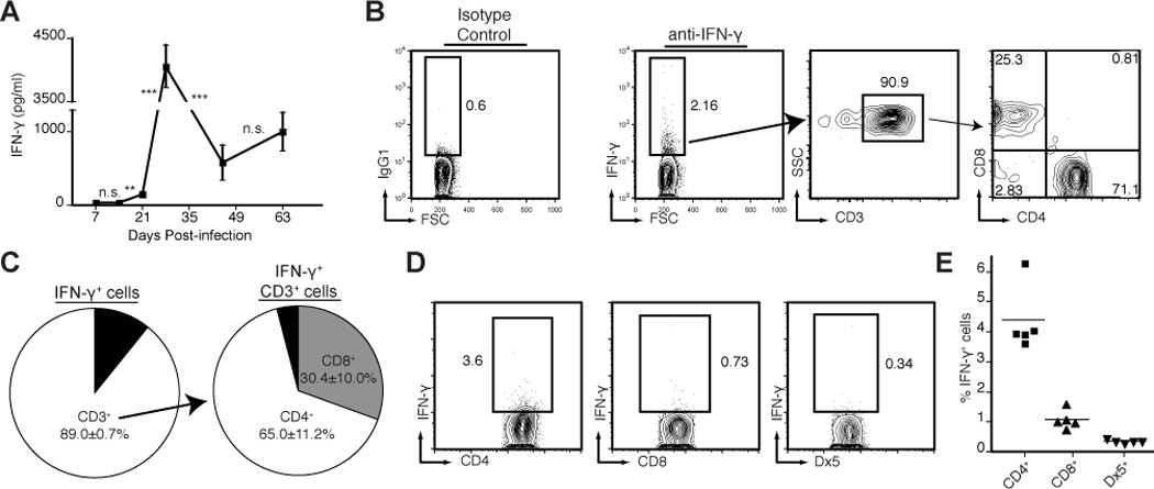
Both CD4+ and CD8+ T cells contribute to immunity to tuberculosis, and both can produce the essential effector cytokine IFN-γ. However, the precise role and relative contribution of each cell type to in vivo IFN-γ production are incompletely understood. To identify and quantitate the cells that produce IFN-γ at the site of Mycobacterium tuberculosis infection in mice, we used direct intracellular cytokine staining ex vivo without restimulation. We found that CD4+ and CD8+ cells were predominantly responsible for production of this cytokine in vivo, and we observed a remarkable linear correlation between the fraction of CD4+ cells and the fraction of CD8+ cells producing IFN-γ in the lungs. In the absence of CD4+ cells, a reduced fraction of CD8+ cells was actively producing IFN-γ in vivo, suggesting that CD4+ effector cells are continually required for optimal IFN-γ production by CD8+ effector cells. Accordingly, when infected mice were treated i.v. with an MHC-II-restricted M. tuberculosis epitope peptide to stimulate CD4+ cells in vivo, we observed rapid activation of both CD4+ and CD8+ cells in the lungs. Indirect activation of CD8+ cells was dependent on the presence of CD4+ cells but independent of IFN-g responsiveness of the CD8+ cells. These data provide evidence that CD4+ cell deficiency impairs IFN-γ production by CD8+ effector cells and that ongoing cross-talk between distinct effector T cell types in the lungs may contribute to a protective immune response against M. tuberculosis. Conversely, defects in these interactions may contribute to susceptibility to tuberculosis and other infections.


Conflict of interest statement

The authors have no conflicting financial interests.

Figures

Figure 1
Figure 1. IFN-γ producing cells in the lungs of M. tuberculosis infected mice
(A) Concentrations of IFN-γ protein in lung homogenates during the initial nine weeks of infection, as measured by IFN-γ ELISA. Asterisks indicate statistical significance of differences in concentration of IFN-γ observed in n = 4 mice, between adjacent time points. ** p<0.005; *** p<0.001. (B) CD3+ T cells account for 90% of the IFN-γ+ cells in the lungs on day 28 post-infection as measured by direct intracellular cytokine staining followed by flow cytometry. IFN-γ-producing cells were identified relative to isotype control antibody staining. Flow cytometry plots show lung cells from a representative mouse. (C) Graphic representation of data from B, showing mean data from n=4 mice. (D) Frequency of IFN-γ producing cells within a given cell subset. Lung cells from day 28 post-infection were gated on relevant markers: CD3+, CD4+; CD3+, CD8+; or CD3, DX5+. (E) Graphic representation of data from D, showing mean data from n=5 mice. All figures represent data from 2 or more experimental replicates. Data are representative of at least 2 independent experiments.
Figure 2
Figure 2. IFN-γ production by CD8+ effector T cells is influenced by CD4+ T cells
(A) IFN-γ production by CD4+ or CD8+ T cells from the lungs of individual mice on day 28 post-infection. Flow cytometry plots show CD3+, CD4+ (top row) or CD8+ (bottom row) cells; values indicate the frequency of IFN-γ+ cells within each population. (B) Linear correlation between the frequency of IFN-γ producing T cells in CD4+ and CD8+ subsets: graphic depiction of data in A, n=5 mice, with linear correlation performed with “x” indicating slope (p = 0.018) and “R2” indicating goodness of fit for linear curve. Data shown in Panels (A) and (B) are from independent experiments. (C) Requirement of CD4+ T cells for IFN-γ production by CD8+ T cells. C57BL/6 or CD4+ T cell deficient MHCII−/− mice were infected with M. tuberculosis and the frequency of IFN-γ+ CD8+ T cells in the lungs was compared, n=4 mice. * p<0.05. Data are representative of at least 2 independent experiments.
Figure 3
Figure 3. Production of IFN-γ by CD8+ T cells requires continual presence of CD4+ T cells
C57BL/6 mice infected with M. tuberculosis were treated with one intraperitoneal dose of monoclonal antibody: either anti-CD4+ or isotype control. Cells were isolated from lungs 24 hours later. The frequency of CD8+ T cells producing IFN-γ in each group was determined by flow cytometry. (A) Flow cytometry plots indicate CD3+ CD8+ cells from the lungs of a representative mouse from either treatment group. Values indicate the frequency of IFN-γ+ cells within each subset 24 hours after treatment. (B) Graphic representation of data from A, indicating the frequency of IFN-γ+ CD8+ T cells in the lungs of n=4 mice. * p<0.05. Data are representative of 2 independent experiments.
Figure 4
Figure 4. Activation of CD4+ effector T cells activates CD8+ effector T cells in vivo
(A) C57BL/6 mice infected with M. tuberculosis were injected intravenously with a synthetic peptide (100 µg) containing a known CD4+ T cell epitope from M. tuberculosis Ag85B, peptide 25 (aa 240–254). 2 hours after injection, the frequency of IFN-γ+ CD4+ or CD8+ T cells was compared to those in infected mice not receiving peptide. Flow cytometry plots show CD3+CD4+ (top row) or CD8+ (bottom row) cells from the lungs of a representative mouse. Values indicate frequency of IFN-γ+ cells in each subset. (B) CD8+ T cell activation upon peptide 25 injection is not due to direct recognition of peptide 25 by CD8+ T cells. Graph depicts the fraction of IFN-γ+ CD8+ T cells in the lungs of either C57BL/6 or MHCII−/− mice, either untreated or 2 hours after injection with 100 µg of either Ag85B peptide 25 (a CD4+ T cell epitope) or Mtb32A309–318 peptide (a CD8+ T cell epitope). Asterisk indicates statistical significance of differences in frequency of IFN-γ+ cells detected among one group of cells from n=4 mice of one genetic background between different treatment groups. * p<0.05; “n.s.”: not significant. Data are representative of 2 independent experiments.
Figure 5
Figure 5. Indirect activation of CD8+ T cells with peptide 25 is independent of IFN-γ signaling
C57BL/6 or IFN-γR−/− mice infected with M. tuberculosis were injected intravenously with Ag85B peptide 25 to activate CD4+ T cells. 2 hours after injection, the frequency of IFN-γ+ T cells was compared to those in infected mice not receiving peptide. Graphs depict the fraction of IFN-γ+ among CD3+, CD4+ (left) or CD3+, CD8+ (right) T cells in the lungs. Asterisks indicate statistical significance of differences in frequency of IFN-γ+ cells detected among one cell type between different treatment groups. * p<0.05; “n.s.”: not significant. Data are representative of 2 independent experiments.

References

    1. Mogues T, Goodrich ME, Ryan L, LaCourse R, North RJ. The relative importance of T cell subsets in immunity and immunopathology of airborne Mycobacterium tuberculosis infection in mice. J Exp Med. 2001;193:271–280. - PMC - PubMed
    1. Muller I, Cobbold SP, Waldmann H, Kaufmann SH. Impaired resistance to Mycobacterium tuberculosis infection after selective in vivo depletion of L3T4+ and Lyt-2+ T cells. Infect Immun. 1987;55:2037–2041. - PMC - PubMed
    1. Filipe-Santos O, Bustamante J, Chapgier A, Vogt G, de Beaucoudrey L, Feinberg J, Jouanguy E, Boisson-Dupuis S, Fieschi C, Picard C, Casanova JL. Inborn errors of IL-12/23- and IFN-gamma-mediated immunity: molecular, cellular, and clinical features. Semin Immunol. 2006;18:347–361. - PubMed
    1. Cooper AM, Dalton DK, Stewart TA, Griffin JP, Russell DG, Orme IM. Disseminated tuberculosis in interferon gamma gene-disrupted mice. J Exp Med. 1993;178:2243–2247. - PMC - PubMed
    1. Flynn JL, Chan J, Triebold KJ, Dalton DK, Stewart TA, Bloom BR. An essential role for interferon gamma in resistance to Mycobacterium tuberculosis infection. J Exp Med. 1993;178:2249–2254. - PMC - PubMed

Publication types

MeSH terms

Substances