. 2012 Aug 30;120(9):1888-98.
doi: 10.1182/blood-2011-09-380808. Epub 2012 Jul 26.

Tyrosine 201 is required for constitutive activation of JAK2V617F and efficient induction of myeloproliferative disease in mice

Affiliations

Tyrosine 201 is required for constitutive activation of JAK2V617F and efficient induction of myeloproliferative disease in mice

Dongqing Yan et al. Blood. .

Abstract

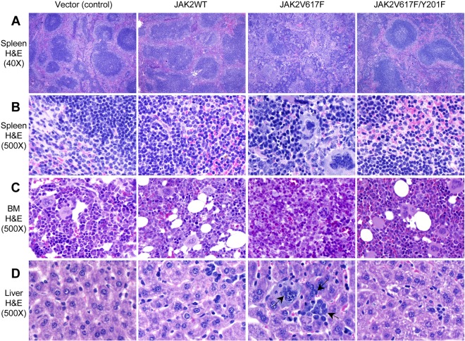
The JAK2V617F mutation has been detected in most cases of Ph-negative myeloproliferative neoplasms (MPNs). The JAK2V617F protein is a constitutively activated tyrosine kinase that leads to transformation of hematopoietic progenitors. Previous studies have shown that several tyrosine residues within JAK2 are phosphorylated on growth factor or cytokine stimulation. However, the role of these tyrosine residues in signaling and transformation mediated by JAK2V617F remains unclear. In this study, we sought to determine the role of tyrosine 201, which is a potential binding site for Src homology 2 domain-containing proteins, in JAK2V617F-induced hematopoietic transformation by introducing a tyrosine-to-phenylalanine point mutation (Y201F) at this site. We observed that the Y201F mutation significantly inhibited cytokine-independent cell growth and induced apoptosis in Ba/F3-EpoR cells expressing JAK2V617F. The Y201F mutation also resulted in significant inhibition of JAK2V617F-mediated transformation of hematopoietic cells. Biochemical analyzes revealed that the Y201F mutation almost completely inhibited constitutive phosphorylation/activation of JAK2V617F. We also show that the Y201 site of JAK2V617F promotes interaction with Stat5 and Shp2, and constitutive activation of downstream signaling pathways. Furthermore, using a BM transduction/transplantation approach, we found that tyrosine 201 plays an important role in the induction of MPNs mediated by JAK2V617F.


Figures

Figure 1
Figure 1
JAK2V617F requires tyrosine 201 for cytokine-independent proliferation and transformation of Ba/F3-EpoR cells. Ba/F3-EpoR cells stably expressing JAK2WT, JAK2V617F, and JAK2V617F/Y201F were used. (A) JAK2 protein expression was assessed in these cell lines by Western blot. (B) Cytokine-independent cell proliferation was determined over 3 days using the WST assay. (C) Total numbers of viable cells were counted over 5 days in the absence of cytokine by trypan blue exclusion. In both cases, JAK2V617F-induced cytokine-independent cell proliferation was significantly inhibited by the Y201F mutation. Results shown are representative of 3 independent experiments. Data are mean ± SEM. (D) Flow cytometric analysis shows a marked increase in apoptosis (annexin V+) in Ba/F3-EpoR cells expressing JAK2V617F/Y201F compared with those expressing JAK2V617F 2 days after cytokine withdrawal. Representative dot plots from 3 independent experiments are shown. (E) Percentages of annexin V+ cells from 3 independent experiments are shown in bar graphs as mean ± SEM. Noticeably, the population undergoing apoptosis (annexin V+) in Ba/F3-EpoR cells expressing JAK2V617F/Y201F was significantly higher than those expressing JAK2V617F and identical to those expressing JAK2WT. (F) The Y201F mutation remarkably inhibited the transformation induced by JAK2V617F. Ba/F3-EpoR cells expressing different JAK2 mutants (2.5 × 10² cells/dish) were plated in duplicate in methylcellulose medium without any cytokine. Colonies were counted after 5 days. Data are mean ± SEM from 3 independent experiments. **P < .005 (1-way ANOVA).
Figure 2
Figure 2
Tyrosine 201 is required for constitutive activation of JAK2V617F and its downstream signaling. (A-C) Ba/F3-EpoR cells expressing JAK2WT, JAK2V617F, and JAK2V617F/Y201F were deprived of cytokine and serum for 6 hours. Tyrosyl phosphorylation of JAK2 (A), Stat5 (B), and Shp2 (C) was detected by immunoprecipitation with specific antibodies against JAK2, Stat5, and Shp2, followed by immunoblotting with phosphotyrosine antibody (4G10). Membranes were reprobed with total antibodies. Note that constitutive tyrosine phosphorylation of JAK2, Stat5, and Shp2 induced by JAK2V617F was significantly inhibited by the Y201F mutation. (D) Ba/F3-EpoR cells expressing JAK2WT, JAK2V617F, and JAK2V617F/Y201F were cytokine and serum-starved followed by stimulation with Epo (3 U/mL) for the indicated times. Cell lysates were prepared and directly immunoblotted with phospho-specific antibodies or total antibodies as indicated. (E) Histograms demonstrate the fold changes in phosphorylation of JAK2, Stat5, Shp2, p70S6K, Akt, and Erk2 compared with the phosphorylation levels in cells expressing JAK2WT. All the data are normalized for the JAK2WT value at 15 minutes, which is set to 1. Data from 3 independent experiments are shown as mean ± SEM. *P < .05 (1-way ANOVA). **P < .005 (1-way ANOVA). Notably, Y201F mutation markedly inhibited the activation of JAK2V617F and downstream signaling pathways, including Stat5, Shp2, p70S6 kinase, Akt, and Erk.
Figure 3
Figure 3
Tyrosine 201 promotes interaction of Stat5 and Shp2 with JAK2V617F. (A-B) JAK2WT, JAK2V617F, or JAK2V617F/Y201F was coexpressed with Stat5A (A) or Shp2 (B) into 293T cells. Stat5A or Shp2 was immunoprecipitated from cell lysates using antibodies against Stat5A (A) or Shp2 (B). Coprecipitated JAK2, Stat5A, and Shp2 were determined by immunoblotting using specific antibodies as indicated. (C-D) Ba/F3-EpoR cells expressing JAK2WT, JAK2V617F, or JAK2V617F/Y201F were cytokine and serum deprived before harvesting. Cell lysates were immunoprecipitated by an anti-Stat5 (C) or anti-Shp2 antibody (D) and subjected to immunoblotting with the indicated antibodies. Note that the Y201F mutation significantly inhibited the interaction between JAK2V617F and Stat5 as well as the interaction between JAK2V617F and Shp2. (E) Y201 is not required for dimerization of JAK2WT or JAK2V617F. The 293T cells were cotransfected with Myc- and HA-tagged JAK2WT or different JAK2 mutants as indicated. Myc-tagged JAK2WT or mutants were efficiently coimmunoprecipitated with HA-tagged JAK2WT or JAK2 mutants. Notably, Y201F mutation did not affect the self-association or dimerization of JAK2WT or JAK2V617F.
Figure 4
Figure 4
Tyrosine 201 is required for efficient induction of MPNs by JAK2V617F. BM from 6-week female Balb/c mice was infected with retroviruses expressing vector (control), JAK2WT, JAK2V617F, or JAK2V617F/Y201F and then transplanted into lethally irradiated Balb/c recipient mice. Peripheral blood red blood cell (A), hemoglobin (B), white blood cell (C), and neutrophil (D) counts in mice receiving JAK2V617F/Y201F-transduced BM (n = 10) were significantly reduced compared with mice receiving JAK2V617F-transduced BM (n = 10) and were comparable with those receiving vector control- or JAK2WT-transduced BM (n = 5) 8 weeks after transplantation. (E) Spleen weight. Mice receiving JAK2V617F-transduced BM developed profound splenomegaly compared with the control (vector or JAK2WT) mice. The Y201F mutation significantly inhibited the JAK2V617F-evoked increase in spleen weight (n = 5 for mice receiving vector control- or JAK2WT-transduced BM; n = 6 for mice receiving JAK2V617F- or JAK2V617F/Y201F-transduced BM). (F) Liver weight. No significant difference was observed in the liver size among all 4 groups of mice (n = 5). (G) Flow cytometric analysis of GFP expression in the BM 8 weeks after transplantation. Histogram represents the percentage of GFP+ population in the BM of the transplanted mice (n = 5). *P < .05 (1-way ANOVA). **P < .005 (1-way ANOVA). Data are mean ± SEM. (H) Southern blot analysis with a radioactive GFP probe demonstrating oligoclonal integration of proviral clones in the BM of the transplanted animals expressing vector (control), JAK2WT, JAK2V617F, or JAK2V617F/Y201F. DNA from Ba/F3-EpoR-JAK2V617F cells is included at right. DNA size markers are shown in kilobases at left.
Figure 5
Figure 5
Histopathologic characterization of the transplanted animals. (A-B) Hematoxylin and eosin stains of the spleens (original magnification ×40 and 500) obtained from vector (control) and JAK2WT expressing mice show normal architecture and minimal extramedullary hematopoiesis. Spleens of JAK2V617F-expressing mice show marked distortion of the splenic architecture with an attenuation of the white pulp and expansion of the red pulp containing abundant extramedullary hematopoiesis, including megakaryocytes and erythroid and granulocyte precursors. JAK2V617F/Y201F-expressing mice show minimal alteration of the architecture with only mild extramedullary hematopoiesis. (C) BM sections (hematoxylin and eosin stain; original magnification ×500) from JAK2V617F-expressing mice show hypercellularity with increased and dysplastic megakaryocytes and increase in granulopoiesis. JAK2V617F/Y201F mouse BM was essentially normal and resembles that of control animals. (D) Liver sections (hematoxylin and eosin stain; original magnification ×500) from JAK2V617F-expressing mice show extramedullary hematopoiesis, whereas livers of JAK2V617F/Y201F mice appear identical to those of control animals. Arrows indicate megakaryocytes and granulocytes in the liver of JAK2V617F mice.
Figure 6
Figure 6
Effects of the Y201F mutation on hematopoietic progenitors expressing JAK2V617F. (A) Flow cytometric analysis demonstrates significant decrease in Ter119+CD71+ erythroid precursors and Gr-1+Mac-1+ myeloid population in the spleens of mice transplanted with JAK2V617F/Y201F-transduced BM compared with mice transplanted with JAK2V617F-transduced BM. Representative dot plots are presented. (B) Histogram shows the percentage of Gr-1+Mac-1+, Ter119+, Ter119+CD71+, and CD71+ populations in the spleens (n = 4 for mice expressing vector control or JAK2WT; n = 6 for mice expressing JAK2V617F; n = 7 for mice expressing JAK2V617F/Y201F). Noticeably, the Y201F mutation markedly inhibited the JAK2V617F-evoked increase in Gr-1+Mac-1+, Ter119+, Ter119+CD71+, and CD71+ populations, and there were no significant differences in those populations among mice transplanted with vector control, JAK2WT-, or JAK2V617F/Y201F-transduced BM. (C) Analysis of Epo-independent CFU-E colony formation in the spleen. A total of 1 × 10⁵ spleen cells were seeded in methylcellulose medium without any cytokine (Methocult M3234). CFU-E colonies were scored after 2 days. Data are mean ± SEM from 3 independent experiments. *P < .05 (1-way ANOVA). **P < .005 (1-way ANOVA).

Similar articles

Cited by

References

    1. James C, Ugo V, Le Couedic JP, et al. A unique clonal JAK2 mutation leading to constitutive signaling causes polycythemia vera. Nature. 2005;434(7037):1144–1148. - PubMed
    1. Levine RL, Wadleigh M, Cools J, et al. Activating mutation in the tyrosine kinase JAK2 in polycythemia vera, essential thrombocythemia, and myeloid metaplasia with myelofibrosis. Cancer Cell. 2005;7(4):387–397. - PubMed
    1. Baxter EJ, Scott LM, Campbell PJ, et al. Acquired mutation of the tyrosine kinase JAK2 in human myeloproliferative disorders. Lancet. 2005;365(9464):1054–1061. - PubMed
    1. Kralovics R, Passamonti F, Buser AS, et al. A gain-of-function mutation of JAK2 in myeloproliferative disorders. N Engl J Med. 2005;352(17):1779–1790. - PubMed
    1. Zhao R, Xing S, Li Z, et al. Identification of an acquired JAK2 mutation in polycythemia vera. J Biol Chem. 2005;280(24):22788–22792. - PMC - PubMed

Publication types

MeSH terms