Review
. 2012 Sep 20;120(12):2376-89.
doi: 10.1182/blood-2012-05-408682. Epub 2012 Jul 26.

How I treat plasma cell leukemia

Affiliations
Review

How I treat plasma cell leukemia

Niels W C J van de Donk et al. Blood. .

Abstract

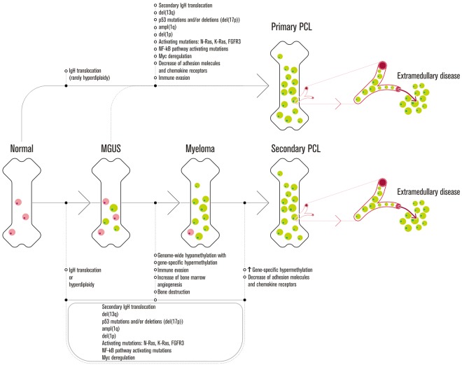
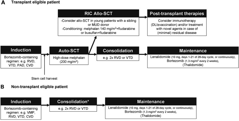
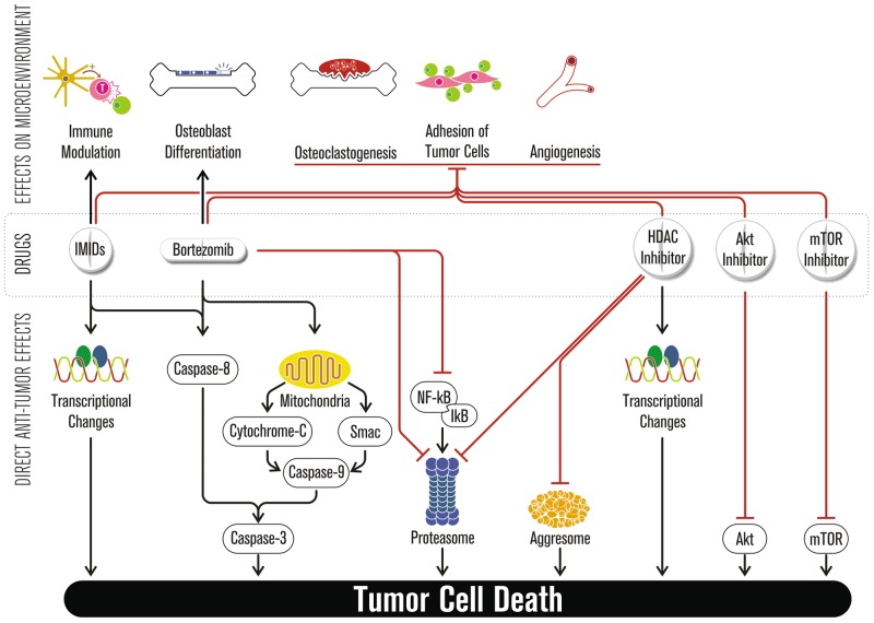
Primary plasma cell leukemia (pPCL) is a rare and aggressive plasma cell proliferative disorder with a very poor prognosis and with distinct biologic, clinical, and laboratory features. Compared with multiple myeloma, pPCL presents more often with extramedullary involvement, anemia, thrombocytopenia, hypercalcemia, elevated serum β(2)-microglobulin and lactate dehydrogenase levels, as well as impaired renal function. Many of the genetic aberrations observed in newly diagnosed pPCL are typically found in advanced multiple myeloma. These cytogenetic abnormalities and mutations lead to increased proliferation, enhanced inhibition of apoptosis, escape from immune surveillance, and independence from the BM microenvironment, with changes in expression of adhesion molecules or chemokine receptors. The outcome of pPCL has improved with the introduction of autologous stem cell transplantation and combination approaches with novel agents, including bortezomib and immunomodulatory drugs, such as lenalidomide. In this review, we provide an overview of currently available therapeutic options with recommendations of how these treatment modalities can best be used to improve outcome for plasma cell leukemia patients.


Figures

Figure 1
Figure 1
Wladyslaw Antoni Gluzinski around 1930. In 1906, Professor A. Gluzinski and Dr M. Reichenstein described the first case of pPCL in the “Wiener Klinische Wochenschrift.”, At that time, the authors worked in the University Hospital of Lemberg (Lvov), which was then part of the Austrian Empire, but is now one of the main cities in western Ukraine. Professor Gluzinski was born in Wloclawek (Poland) in 1856 and died in Warsaw in 1935. The authors describe a 47-year-old train ticket inspector who presented with bone pain, a palpable mass over one of his ribs, rib fractures, anemia, and splenomegaly. Urine contained large amounts of a proteinaceous substance. In the blood smear, the authors observed erythroblasts and immature plasma cells, sometimes with 2 or 3 nuclei. The absolute number of circulating tumor cells increased significantly during further follow-up (one week before patient died, leucocytes were estimated at 39.4 × 10⁹/L, containing 91% plasma cells). The diagnosis of MM and “leucaemia lymphatica plasmocellularis” was made. Treatment consisted of the organoarsenic compounds Atoxyl and Neoarsicodile. This was followed by a brief period with reduced levels of pain and reduction of the size of the rib tumor, enabling him to resume working. However, the pain and rib tumors recurred and the patient died 6 months after his first presentation of pneumonia. Autopsy confirmed the premortem diagnosis. Interestingly, much later, several groups reported activity from arsenic in MM and related disorders.
Figure 2
Figure 2
Model for the mechanisms that contribute to the development of primary and secondary plasma cell leukemia. Primary immunoglobulin heavy chain (IgH) translocations with 5 recurrent chromosomal partners (4p16, 6p21, 11q13, 16q23, and 20q11) and hyperdiploidy (typically with trisomies of chromosomes 3, 5, 7, 9, 11, 15, 19, and 21) are early events and associated with the initiation of limited clonal plasma cell proliferation in MGUS. Acquisition of secondary chromosomal abnormalities (such as deletions of [parts of] chromosomes or secondary chromosomal translocations) and mutations involving individual genes results in the stepwise progression from MGUS to newly diagnosed symptomatic MM and, finally, aggressive forms of MM, such as sPCL or extramedullary MM. During this process, there is a progressive replacement of normal/polyclonal plasma cells (red) by clonal plasma cells (green). Progression of the plasma cell disorder is also accompanied by altered interactions of the tumor cells with various components of their microenvironment, such as osteoclasts, endothelial cells, and cells of the immune system. On the other hand, the immortalized cell may accumulate various genetic aberrations in a short period of time, resulting in de novo pPCL or rapid progression of MGUS to PCL, without preceding MM. Importantly, in pPCL, hyperdiploidy is very uncommon, whereas primary IgH translocations have a higher incidence compared with the incidence of these primary genetic events in newly diagnosed MM.
Figure 3
Figure 3
Peripheral blood and BM features of pPCL. (A) May-Grünwald-Giemsa staining of peripheral blood smears from 3 different patients with pPCL. (i) The peripheral blood smear of patient 1 is characterized by the presence of circulating plasma cells, including binucleated cells. Some plasma cells have a lymphoplasmacytoid appearance. There is also rouleaux formation and leuko-erythroblastosis (myelocyte in Ai). Cytogenetic analysis demonstrated the presence of an IgH translocation with an unknown partner chromosome as well as deletion of the Y chromosome. (ii) Peripheral blood of patient 2 contains circulating plasma cells, some of which have 2 nuclei or a lymphoplasmacytoid aspect. FISH analysis showed the presence of t(14;16) and ampl(1q21). (iii) The staining of patient 3 shows very atypical circulating plasma cells often with a monocytic appearance. Numerous cells contain cytoplasmic (Russell bodies) and intranuclear inclusions (Dutcher bodies). Cytogenetic analysis revealed multiple deletions, including chromosomes 6q, 12p, 13, 16, and 20, and the presence of t(14;20). (B) May-Grünwald-Giemsa-stained BM smears from the same 3 pPCL (i-iii) patients. (C) Immunophenotypic analysis of the leukemic plasma cells from the patient shown in subpanels Aii and Bii. The tumor cells are positive for CD138 and CD27, weakly positive for CD38 and CD45, and negative for CD56 and CD19. Cells were also negative for CD20 (not shown).
Figure 4
Figure 4
Treatment of pPCL. Flow chart with suggestions for how to treat patients with pPCL. *Consolidation for non–transplant-eligible patients is given once best response to induction therapy is achieved. Varicella zoster virus prophylaxis with acyclovir or valacyclovir is recommended for all patients receiving bortezomib-based therapy. Thromboprophylaxis (aspirin for patients at standard risk for thromboembolic events and low-molecular-weight heparin or adjusted-dose warfarin in high-risk patients) is indicated when either thalidomide or lenalidomide is combined with dexamethasone or chemotherapy. MUD indicates matched unrelated donor; DLI, donor lymphocyte infusion; PAD, bortezomib, adriamycin, and dexamethasone; CVD, cyclophosphamide, bortezomib, and dexamethasone; VMP, bortezomib, melphalan, and prednisone; and RIC, reduced intensity conditioning.
Figure 5
Figure 5
Novel agents in plasma cell leukemia. Immunomodulatory drugs, bortezomib, and next-generation novel agents have direct antitumor effects but also affect the BM microenvironment (ie, down-regulation of adhesion molecules, reduction of osteoclastogenesis, promotion of new bone formation, inhibition of angiogenesis, and stimulation of immune effector cells).

Similar articles

Cited by

References

    1. Kyle RA, Maldonado JE, Bayrd ED. Plasma cell leukemia: report on 17 cases. Arch Intern Med. 1974;133(5):813–818. - PubMed
    1. International Myeloma Working Group. Criteria for the classification of monoclonal gammopathies, multiple myeloma and related disorders: a report of the International Myeloma Working Group. Br J Haematol. 2003;121(5):749–757. - PubMed
    1. Gluzinski A, Reichenstein M. Myeloma und leucaemia lymphatica plasmocellularis. Wien Klin Wochenschr. 1906;12:336–339.
    1. Avet-Loiseau H, Roussel M, Campion L, et al. Cytogenetic and therapeutic characterization of primary plasma cell leukemia: the IFM experience. Leukemia. 2012;26(1):158–159. - PubMed
    1. Tiedemann RE, Gonzalez-Paz N, Kyle RA, et al. Genetic aberrations and survival in plasma cell leukemia. Leukemia. 2008;22(5):1044–1052. - PMC - PubMed

MeSH terms