. 2012 Jul 25;4(144):144ra102.
doi: 10.1126/scitranslmed.3003875.

Temsirolimus activates autophagy and ameliorates cardiomyopathy caused by lamin A/C gene mutation

Affiliations

Temsirolimus activates autophagy and ameliorates cardiomyopathy caused by lamin A/C gene mutation

Jason C Choi et al. Sci Transl Med. .

Abstract

Mutations in the lamin A/C gene (LMNA), which encodes A-type lamins, cause a diverse range of diseases collectively called laminopathies, the most common of which is dilated cardiomyopathy. Emerging evidence suggests that LMNA mutations cause disease by altering cell signaling pathways, but the specific mechanisms are poorly understood. We show that the AKT-mammalian target of rapamycin pathway is hyperactivated in hearts of mice with cardiomyopathy caused by Lmna mutation and that in vivo administration of the rapamycin analog temsirolimus prevents deterioration of cardiac function. We also show defective autophagy in hearts of these mice and demonstrate that improvement in heart function induced by pharmacological interventions is correlated with enhanced autophagy. These findings provide a rationale for treatment of LMNA cardiomyopathy with rapalogs and implicate defective autophagy as a pathogenic mechanism of cardiomyopathy arising from LMNA mutation.


Conflict of interest statement

Competing interests: H.J.W. and A.M. are inventors on a pending patent application (PCT/US09/42614) on methods for treating and/or preventing cardiomyopathies by ERK and JNK inhibition filed by the Trustees of Columbia University in the City of New York. H.J.W and J.C.C. are inventors on a provisional patent application on the use of rapamycin and rapamycin analogs for the treatment of dilated cardiomyopathies currently planned for filing by the Trustees of Columbia University in the City of New York.

Figures

Fig. 1
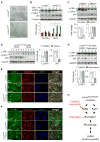
Fig. 1. AKT/mTORC1 is activated in LMNA cardiomyopathy
(A) Western blot analysis of phospho-serine 473 AKT [(pAKT(S473)], phospho-threonine 308 AKT [pAKT(T308)], total AKT, phosphorylated mTOR (pmTOR), total mTOR, phosphorylated S6 (pS6), and total S6 in hearts of 4 to 16 week-old LmnaH222P/H222P (LmnaH222P) and wild-type (LmnaWT) mice (top). Numbers on top of blots denote individual heart samples. Representative blots from two independent experiments. Quantification of pmTOR, pAKT, and pS6 normalized to total mTOR, AKT, and S6, respectively, presented as fold-change over LmnaWT (bottom); n = 4. (B) Western blot of pmTOR, mTOR, pAKT(S473), pAKT(T308), and total AKT, in hearts of 20 week-old LmnaH222P mice treated with DMSO or selumetinib for 4 weeks (top). Numbers on top of blots denote individual heart samples. Quantification of pmTOR and pAKT normalized to total mTOR and AKT, respectively, presented as fold-change over DMSO (bottom); n = 6. (C) Western blot of pAKT (S473 and T308), total AKT and α-tubulin in hearts from human subjects with (LMNAmt) or without (LMNAwt) LMNA cardiomyopathy (top). Numbers on top of blots denote individual heart samples. Quantification of S473 and T308 pAKT normalized to total AKT, presented as fold-change over LMNAwt (bottom); n = 3.
Fig. 2
Fig. 2. mTORC1 inhibition ameliorates cardiomyopathy in LmnaH222P/H222P mice
(A) Western blot of pmTOR, total mTOR, pS6, and total pS6 in hearts of 16 week-old LmnaH222P/H222P mice treated with DMSO or temsirolimus (Temsir) for two weeks (left). Numbers on top of blots denote individual heart samples. Quantification of pmTOR and pS6 normalized to total mTOR and S6, respectively, presented as fold-change over DMSO (right); n = 5. (B) Representative hearts of 16 week-old LmnaH222P/H222P mice treated DMSO or temsirolimus. Scale bar = 1 cm. (C) Representative M-mode echocardiographic tracings of 16 week-old LmnaH222P/H222P mice treated with DMSO or temsirolimus. (D) Graphic representations and table with numerical values for left ventricular end diastolic diameter (LVEDD), left ventricular end systolic diameter (LVESD) and fractional shortening (FS) of hearts of 16 week-old LmnaH222P/H222P mice treated with DMSO or temsirolimus; n = 8. (E) qPCR analysis on NppA, NppB, Col1a1, Col1a2 and Fn1 mRNAs in hearts of 16 week-old LmnaH222P/H222P mice treated with DMSO or temsirolimus. Tnni3 mRNA used as internal control. n = 5. (F) Western blot of atrial natriuretic peptide (ANP) and GAPDH in hearts of 16 week-old LmnaH222P/H222P mice treated with DMSO or temsirolimus (top). Numbers on top of blots denote individual heart samples. Quantification of ANP normalized to GAPDH presented as fold-change over DMSO (bottom); n = 5.
Fig. 3
Fig. 3. Autophagy is impaired in hearts of LmnaH222P/H222P mice
(A) Western blot of non-lipidated LC3B (LC3B-I), lipidated LC3B (LC3B-II), p62, beclin1, and GAPDH in hearts of 4 to 16 week-old LmnaH222P/H222P (LmnaH222P) and wild-type (LmnaWT) mice (left). Numbers on top of blots denote individual mouse heart samples. (B) Quantification of LC3B-II and p62 normalized to GAPDH presented as fold-change over LmnaWT (right); n = 6; *p= 0.03; **p=0.002; #p=0.04. (C) Western blot of LC3B-I, LC3B-II, p62 and α-tubulin in hearts of 16 week-old LmnaH222P and LmnaWT mice treated with chloroquine (Chloroq) or vehicle (Ctrl) for 10 days (top). Numbers on top of blots denote individual heart samples. Quantification of LC3B-II and p62 normalized to α-tubulin presented as fold-change over Ctrl (bottom); n = 3. (D) Western blot of LC3B-I, LC3B-II, p62 and α-tubulin in hearts of 16 week-old LmnaH222P and LmnaWT mice fasted for 24 hr (Fasted) or fed ad libitum (Fed) (top). Numbers on top of blots denote individual heart samples. Quantification of LC3B-II and p62 normalized to α-tubulin presented as fold-change over Fed (bottom); n = 3.
Fig. 4
Fig. 4. Reactivation of autophagy is associated with improved cardiac function
(A) Photomicrographs of cardiomyocytes from 12 week-old LmnaH222P/H222P(LmnaH222P) and wild-type (LmnaWT) mice; scale bar = 120 μM. (B) Representative Western blot of LC3B-I, LC3B-II, p62 and α-tubulin in cardiomyocytes from LmnaH222P and LmnaWT mice deprived of glucose (Gluc -) for 0, 2 and 4 hr (top). Quantification of LC3B-II and p62 normalized to α-tubulin presented as fold-change over LmnaWT (bottom); n = 3; *p= 0.011; #p=0.046. (C) Western blot of LC3B-I, LC3B-II, p62 and GAPDH in hearts from human subjects with (LMNAmt) or without (LMNAwt) LMNA cardiomyopathy (top). Numbers on top of blots denote individual samples. Quantification of LC3B-II and p62 normalized to α-tubulin presented as fold-change over LMNAwt (bottom); n = 3. (D) Western blot of LC3B-I, LC3B-II, p62, and α-tubulin in hearts of 16 week-old LmnaH222P/H222P mice treated with DMSO or temsirolimus (Temsir) (left). Numbers on top of blots denote individual heart samples. Quantification of LC3B-II and p62 normalized to α-tubulin presented as fold-change over DMSO (right); n = 5. (E) Immunofluorescence micrographs of heart sections from 16 week-old LmnaWT, DMSO-treated LmnaH222P and temsirolimus-treated LmnaH222P mice probed with antibodies against α-actin and ubiquitin. 4’,6-diamidino-2-phenylindole (DAPI) was used as a nuclear counterstain. Representative figures from two independent hearts per group are shown. Scale bar = 30 μM. (F) Immunofluorescence micrographs of heart sections from 16 week-old LmnaWT, DMSO-treated LmnaH222P and temsirolimus-treated LmnaH222P mice probed with antibodies against α-actin and lamin A/C. DAPI was used as a nuclear counterstain. Representative figures from two independent hearts per group are shown. Scale bar = 30 μM. (G) Representative Western blot of LC3B-I, LC3B-II, p62, and GAPDH in hearts of 20 week-old LmnaH222P/H222P mice treated with DMSO or selumetinib (top). Numbers on top of blots denote individual heart samples. Quantification of LC3B-II and p62 normalized to α-tubulin presented as fold-change over DMSO (bottom); n = 6. (H) Schematic of the proposed pathogenesis of LMNA cardiomyopathy. See text for details.

References

    1. Dauer WT, Worman HJ. The nuclear envelope as a signaling node in development and disease. Dev Cell. 2009;17:626–638. - PubMed
    1. Lu JT, Muchir A, Nagy PL, Worman HJ. LMNA cardiomyopathy: cell biology and genetics meet clinical medicine. Dis Model Mech. 2011;4:562–568. - PMC - PubMed
    1. Hernandez L, Roux KJ, Wong ES, Mounkes LC, Mutalif R, Navasankari R, Rai B, Cool S, Jeong JW, Wang H, Lee HS, Kozlov S, Grunert M, Keeble T, Jones CM, Meta MD, Young SG, Daar IO, Burke B, Perantoni AO, Stewart CL. Functional coupling between the extracellular matrix and nuclear lamina by Wnt signaling in progeria. Dev Cell. 2010;19:413–425. - PMC - PubMed
    1. Cao K, Graziotto JJ, Blair CD, Mazzulli JR, Erdos MR, Krainc D, Collins FS. Rapamycin reverses cellular phenotypes and enhances mutant protein clearance in hutchinson-gilford progeria syndrome cells. Sci Transl Med. 2011;3:89ra58. - PubMed
    1. Muchir A, Pavlidis P, Decostre V, Herron AJ, Arimura T, Bonne G, Worman HJ. Activation of MAPK pathways links LMNA mutations to cardiomyopathy in Emery-Dreifuss muscular dystrophy. J Clin Invest. 2007;117:1282–1293. - PMC - PubMed

Publication types