

Spontaneous, uncomplicated dissolution of a large cotton fiber in the laser in situ keratomileusis interface
- PMID: 22837633
- PMCID: PMC3401809
- DOI: 10.4103/0974-9233.97960
Spontaneous, uncomplicated dissolution of a large cotton fiber in the laser in situ keratomileusis interface
Abstract
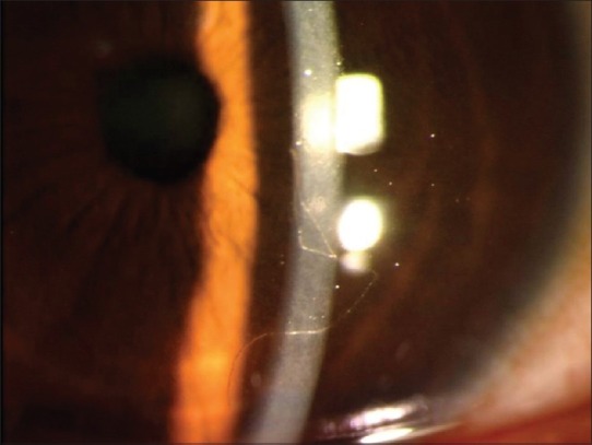
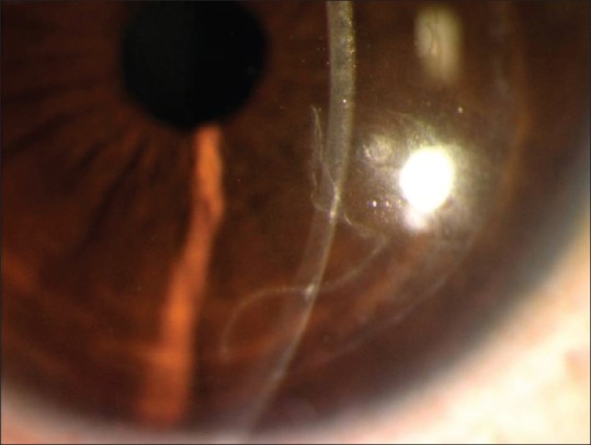
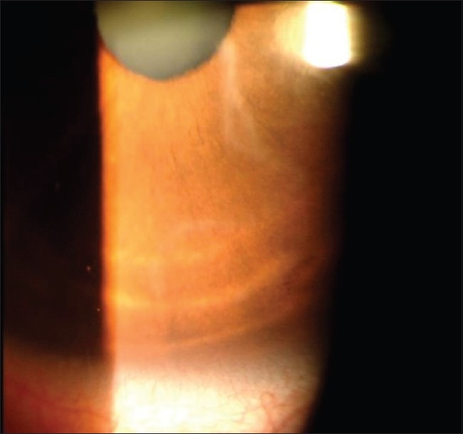
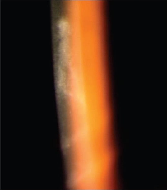
In this article, we report the spontaneous dissolution of a long cotton thread in the laser in situ keratomileusis (LASIK) flap interface. In this observational case report, sequential follow-up of a post-LASIK eye with a long cotton fiber noticed in the LASIK interface was performed. The postoperative course was uneventful, with no evidence of infection, uveitis, or any other complications. The cotton thread underwent spontaneous dissolution. Conservative management of a cotton fiber, not causing any symptoms and noticed after the immediate postoperative follow-up period is over, seems to be a possible alternative to flap relift and intervention.
Keywords: Fiber; Laser In Situ Keratomileusis Interface; Resolution.
Conflict of interest statement
Figures
References
-
- Eisemann J, Carkeet A, Swann PG. Large interface particles from LASIK surgery. Clin Exp Optom. 2006;89:253–6. - PubMed
-
- Crowther KS, Ellingham RB. Complicated removal of corneal foreign bodies 18 months after laser in situ keratomileusis. J Cataract Refract Surg. 2005;31:851–2. - PubMed
-
- Ivarsen A, Thogersen J, Keiding SR, Hjortdal JO, Moller-Pedersen T. Plastic particles at the LASIK interface. Ophthalmology. 2004;111:18–23. - PubMed
-
- Porges Y, Landau D, Douieb J, Levinger S. Removal of corneal foreign bodies following laser in situ keratomileusis. J Refract Surg. 2001;17:559–60. - PubMed
Publication types
MeSH terms
LinkOut - more resources
Full Text Sources
