Clinical Trial
. 2012 Jul 28;14(1):50.
doi: 10.1186/1532-429X-14-50.

Left ventricular remodeling and hypertrophy in patients with aortic stenosis: insights from cardiovascular magnetic resonance

Affiliations
Clinical Trial

Left ventricular remodeling and hypertrophy in patients with aortic stenosis: insights from cardiovascular magnetic resonance

Marc R Dweck et al. J Cardiovasc Magn Reson. .

Abstract

Background: Cardiovascular magnetic resonance (CMR) is the gold standard non-invasive method for determining left ventricular (LV) mass and volume but has not been used previously to characterise the LV remodeling response in aortic stenosis. We sought to investigate the degree and patterns of hypertrophy in aortic stenosis using CMR.

Methods: Patients with moderate or severe aortic stenosis, normal coronary arteries and no other significant valve lesions or cardiomyopathy were scanned by CMR with valve severity assessed by planimetry and velocity mapping. The extent and patterns of hypertrophy were investigated using measurements of the LV mass index, indexed LV volumes and the LV mass/volume ratio. Asymmetric forms of remodeling and hypertrophy were defined by a regional wall thickening ≥ 13 mm and >1.5-fold the thickness of the opposing myocardial segment.

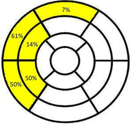
Results: Ninety-one patients (61 ± 21 years; 57 male) with aortic stenosis (aortic valve area 0.93 ± 0.32 cm2) were recruited. The severity of aortic stenosis was unrelated to the degree (r2=0.012, P=0.43) and pattern (P=0.22) of hypertrophy. By univariate analysis, only male sex demonstrated an association with LV mass index (P=0.02). Six patterns of LV adaption were observed: normal ventricular geometry (n=11), concentric remodeling (n=11), asymmetric remodeling (n=11), concentric hypertrophy (n=34), asymmetric hypertrophy (n=14) and LV decompensation (n=10). Asymmetric patterns displayed considerable overlap in appearances (wall thickness 17 ± 2mm) with hypertrophic cardiomyopathy.

Conclusions: We have demonstrated that in patients with moderate and severe aortic stenosis, the pattern of LV adaption and degree of hypertrophy do not closely correlate with the severity of valve narrowing and that asymmetric patterns of wall thickening are common.

Trial registration: ClinicalTrials.gov NCT00930735.


Figures

Figure 1
Figure 1
CMR definitions of the six patterns of left ventricular hypertrophy and remodeling in aortic stenosis. Schematic representation of the left ventricular structure alongside CMR short-axis images of the left ventricle in end-diastole. Normal ventricular structure: characterised by a normal LV mass index, indexed LVEDV, and a normal M/V. Concentric remodeling: characterised by an increased M/V and normal LV mass index. Asymmetric remodeling: similar to concentric remodeling except that in addition there is evidence of asymmetric wall thickening. Concentric Hypertrophy: characterised by an increased M/V and LV mass index. Asymmetric hypertrophy: similar to concentric hypertrophy except that in addition there is evidence of asymmetric wall thickening.Left Ventricular Decompensation: characterised by a dilated left ventricle and normal M/V. The LV mass index may be increased primarily due to LV dilatation. Note no patients fulfilled the criteria for eccentric hypertrophy and so this was replaced by LV decompensation. increased; decreased; = normal; ✓present; ✖absent.
Figure 2
Figure 2
Lack of correlation between aortic valve area and left ventricular mass index.A. Total population. B. Population after excluding patients with hypertension. C. Males. D. Females.
Figure 3
Figure 3
Site of maximal wall thickening in asymmetric hypertrophy and remodeling based on the 17-segment model of the left ventricle. Asymmetric wall thickening was observed in the basal anterior wall in 7%, otherwise it was confined to the septum at the basal and mid-cavity levels.

References

    1. Carabello BA. The relationship of left ventricular geometry and hypertrophy to left ventricular function in valvular heart disease. J Heart Valve Dis. 1995;4(Suppl 2):S132–S138. discussion S138–139. - PubMed
    1. Grossman W, Jones D, McLaurin L. Wall stress and patterns of hypertrophy in the human left ventricle. J Clin Invest. 1975;56:56–64. doi: 10.1172/JCI108079. - DOI - PMC - PubMed
    1. Gradman AH, Alfayoumi F. From left ventricular hypertrophy to congestive heart failure: management of hypertensive heart disease. Prog Cardiovasc Dis. 2006;48:326–341. doi: 10.1016/j.pcad.2006.02.001. - DOI - PubMed
    1. Gosse P. Left ventricular hypertrophy as a predictor of cardiovascular risk. J Hypertens Suppl. 2005;23:S27–S33. doi: 10.1097/01.hjh.0000165625.79933.9a. - DOI - PubMed
    1. Cioffi G, Faggiano P, Vizzardi E, Tarantini L, Cramariuc D, Gerdts E, de Simone G. Prognostic effect of inappropriately high left ventricular mass in asymptomatic severe aortic stenosis. Heart. 2011;97:301–307. doi: 10.1136/hrt.2010.192997. - DOI - PubMed

Publication types

MeSH terms

Associated data