. 2012 Dec 10;21(18):3289-97.
doi: 10.1089/scd.2012.0095. Epub 2012 Sep 28.

Microvesicles derived from human umbilical cord mesenchymal stem cells stimulated by hypoxia promote angiogenesis both in vitro and in vivo

Affiliations

Microvesicles derived from human umbilical cord mesenchymal stem cells stimulated by hypoxia promote angiogenesis both in vitro and in vivo

Hong-Chao Zhang et al. Stem Cells Dev. .

Abstract

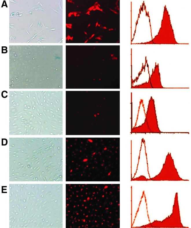
Although mesenchymal stem cells (MSCs) have been increasingly trialed to treat a variety of diseases, the underlying mechanisms remain still elusive. In this study, human umbilical cord (UC)-derived MSCs were stimulated by hypoxia, and the membrane microvesicles (MVs) in the supernatants were collected by ultracentrifugation, observed under an electron microscope, and the origin was identified with the flow cytometric technique. The results showed that upon hypoxic stimulus, MSCs released a large quantity of MVs of ~100 nm in diameter. The MVs were phenotypically similar to the parent MSCs, except that the majority of them were negative for the receptor of platelet-derived growth factor. DiI-labeling assay revealed that MSC-MVs could be internalized into human UC endothelial cells (UC-ECs) within 8 h after they were added into the culture medium. Carboxyfluorescein succinimidyl ester-labeling technique and MTT test showed that MSC-MVs promoted the proliferation of UC-ECs in a dose-dependent manner. Further, MVs could enhance in vitro capillary network formation of UC-ECs in a Matrigel matrix. In a rat hindlimb ischemia model, both MSCs and MSC-MVs were shown to improve significantly the blood flow recovery compared with the control medium (P<0.0001), as assessed by laser Doppler imaging analysis. These data indicate that MV releasing is one of the major mechanisms underlying the effectiveness of MSC therapy by promoting angiogenesis.


Figures

FIG. 1.
FIG. 1.
Umbilical cord mesenchymal stem cells (UC-MSCs) shed microvesicles (MVs). (A) Representative flow cytometry results show that cells in serum-free medium (SF) under hypoxia (Hypo) were inclined to apoptosis compared with the cells normoxia (Norm). X-axis represents Annexin-V-fluorescent isothiocyanate, and Y-axis indicates propidium iodide (PI). (B) Morphological features of MVs under electron microscope; bar: 100 nm. (C) Identification of MSC-MVs by flow cytometry. Upper panel: washed beads (left) or MV-coated beads after reaction with isotype antibodies (right) were collected, and the single beads (green) and doublets (pink red) were gated for fluorescence analysis. MVs: MSC-MVs, MSCs: the parent MSCs. X-axis: forward scatter corner (FSC). Y-axis: phycoerythrin-conjugated antibodies against the antigens of choice. The positive and negative proportions are indicated. The results are representative of 2 individual experiments. Color images available online at www.liebertpub.com/scd
FIG. 2.
FIG. 2.
The fluorescence of DiI-labeled MSCs and MSC-MV-treated endothelial cells (ECs). MSCs were labeled with DiI dye (A) and umbilical cord ECs (UC-ECs) were cultured in normoxia (B, D) or hypoxia (C, E) in the presence of DiI-MVs for 2 h (B, C) and 8 h (D, E). The photographs were taken under an ordinary light (left column) or a fluorescent light (middle column). The typical histograms from flow cytometric tests are also indicated (right column). In the histograms, X-axis indicates the relative fluorescence of DiI, and Y-axis represents number of events; the hollow histograms represent the controls, and the solid ones indicate the tested cells. The results are representative of 2 separate experiments. Color images available online at www.liebertpub.com/scd
FIG. 3.
FIG. 3.
MSC-MVs promoted the proliferation of UC-ECs. (A) Carboxyfluorescein succinimidyl ester (CFSE)-labeled human UC-ECs from 3 samples were cultured in the presence of MSC-MVs at graded concentrations for 72 h. The proportions of the cells that had experienced different divisions were analyzed by flow cytometry technique. Cells that were maintained in 1% fetal bovine serum (FBS) were served as sham controls (ctr) and those in the presence of 5% FBS and 10% FBS were served as positive controls. Three main portions are indicated, including histograms with the colors of pink (generation 2), white blue (generation 3), and yellow (generation 4). X-axis represents CFSE intensity, and Y-axis represents the number of the events. (B) MTT assay shows the stimulation effect of MSC-MVs on the proliferation of UC-ECs. Y-axis: optical density at a wavelength of 490 nm. The results here were from ECs of 2 subjects. Color images available online at www.liebertpub.com/scd
FIG. 4.
FIG. 4.
MSC-MVs promoted tube formation of UC-ECs in a dose-dependent manner. ECs were incubated in Matrigel in the presence of graded doses of MSC-MVs in hypoxia or normoxia. Typical morphology of the tube-like structure is shown in rows (A) (hypoxia) and (B) (normoxia). (C) Y-axis represents the structure number per field, and X-axis indicates different groups. The results are representative of those from 2 individual experiments.
FIG. 5.
FIG. 5.
Representative Laser Doppler images of the rat hindlimbs. Laser Doppler blood flow was performed 2 days after the artery ligation (ctr) without any treatment or 2 weeks after operation (groups as indicated). The colors that correspond to the blood perfusion are shown. Color images available online at www.liebertpub.com/scd
FIG. 6.
FIG. 6.
Typical histological changes of the adductor muscles. (A) Inflammatory granulomatosis; (B) muscle fiber bunches. Arrowhead: small vessels; Arrow: capillaries. Bar: 25 μm. Color images available online at www.liebertpub.com/scd

References

    1. Pittenger MF. Mackay AM. Beck SC. Jaiswal RK. Douglas R. Mosca JD. Moorman MA. Simonetti DW. Craig S. Marshak DR. Multilineage potential of adult human mesenchymal stem cells. Science. 1999;284:143–147. - PubMed
    1. Dominici M. Le Blanc K. Mueller I. Slaper-Cortenbach I. Marini F. Krause D. Deans R. Keating A. Prockop D. Horwitz E. Minimal criteria for defining multipotent mesenchymal stromal cells. The international society for cellular therapy position statement. Cytotherapy. 2006;8:315–317. - PubMed
    1. Caplan AI. Adult mesenchymal stem cells for tissue engineering versus regenerative medicine. J Cell Physiol. 2007;213:341–347. - PubMed
    1. Short BJ. Brouard N. Simmons PJ. Prospective isolation of mesenchymal stem cells from mouse compact bone. Methods Mol Biol. 2009;482:259–268. - PubMed
    1. Meirelles LS. Chagastelles PC. Nardi NB. Mesenchymal stem cells reside in virtually all post-natal organs and tissues. J Cell Sci. 2006;119:2204–2213. - PubMed

Publication types

Substances

LinkOut - more resources