. 2012 Aug 21;51(33):6584-94.
doi: 10.1021/bi300876h. Epub 2012 Aug 9.

Nucleotide release sequences in the protein kinase SRPK1 accelerate substrate phosphorylation

Affiliations

Nucleotide release sequences in the protein kinase SRPK1 accelerate substrate phosphorylation

Brandon E Aubol et al. Biochemistry. .

Abstract

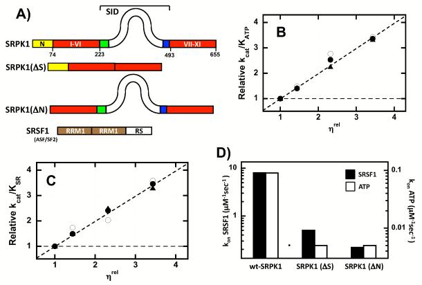
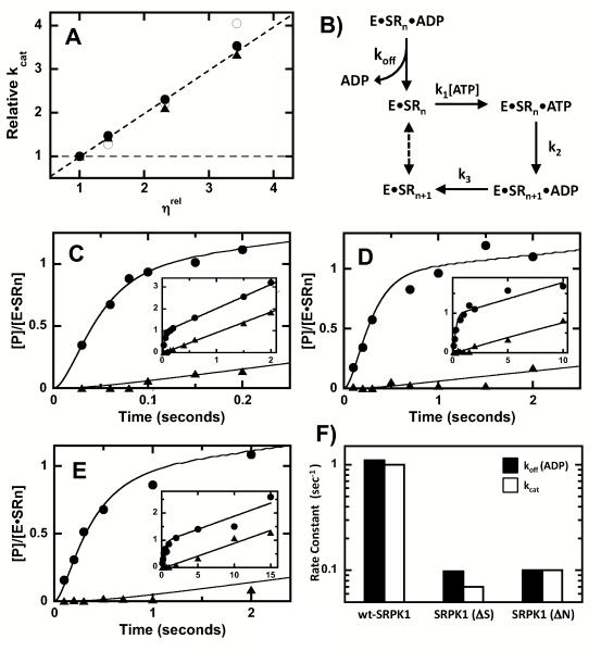
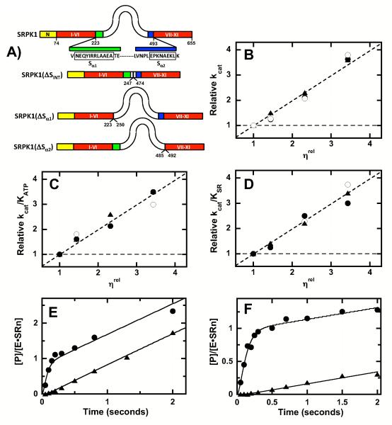
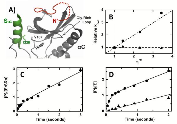
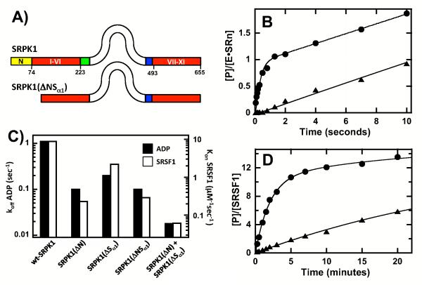
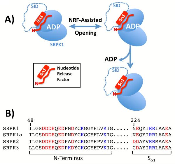
Protein kinases are essential signaling enzymes that transfer phosphates from bound ATP to select amino acids in protein targets. For most kinases, the phosphoryl transfer step is highly efficient, while the rate-limiting step for substrate processing involves slow release of the product ADP. It is generally thought that structural factors intrinsic to the kinase domain and the nucleotide-binding pocket control this step and consequently the efficiency of protein phosphorylation for these cases. However, the kinase domains of protein kinases are commonly flanked by sequences that regulate catalytic function. To address whether such sequences could alter nucleotide exchange and, thus, regulate protein phosphorylation, the presence of activating residues external to the kinase domain was probed in the serine protein kinase SRPK1. Deletion analyses indicate that a small segment of a large spacer insert domain and a portion of an N-terminal extension function cooperatively to increase nucleotide exchange. The data point to a new mode of protein kinase regulation in which select sequences outside the kinase domain constitute a nucleotide release factor that likely interacts with the small lobe of the kinase domain and enhances protein substrate phosphorylation through increases in ADP dissociation rate.


Figures

Figure 1
Figure 1. Diffusion Limits of ATP and SRSF1 in the Active Site of Wild-Type & Mutant Forms of SRPK1
A) Enzyme & Substrate Constructs. B,C) Viscosity Effects on Steady-State Kinetic Parameters. Relative values for kcat/KATP (B), and kcat/KSR (C) in the absence and presence of varying sucrose concentrations are measured as a function of relative solvent viscosity (ηrel) for wild-type SRPK1 (○), SRPK1(ΔN) (•) and SRPK1(ΔS) (▲). Dotted lines reflect theoretical slope limits of 0 and 1. D) Bar graph showing changes in the association rate constants for SRSF1 and ATP.
Figure 2
Figure 2. N-Terminus & SID Promote ADP Dissociation from SRPK1
A) Viscosity effects on kcat for wild-type SRPK1 (○), SRPK1(ΔN) (•) and SRPK1(ΔS) (▲). Dotted lines reflect theoretical slope limits of 0 and 1. B) Kinetic Mechanism. Enzyme (E) and SRSF1 (SRn) are preequilibrated in the absence or presence of ADP and the reaction is started with ATP (k1). SRSF1 (SRn) is phosphorylated at the first site (k2), ADP is released (k3) and the product (SRn+1) becomes the substrate for the next phosphorylation event (dotted line). C-E) CATTRAP Experiments. SRSF1 and wild-type SRPK1 (C), SRPK1(ΔN) (D) and SRPK1(ΔS) (E) are prequilibrated in the absence (•) and presence (▲) of 120 ΔM ADP (in syringe) before the reaction is initiated with 600 ΔM 32P-ATP. Insets show extended time frames for the reactions (2-15 sec). The data are simulated to obtain values for koff, k2, k3 and [E*SRn] (Table 2). F) Bar graph showing changes in koff for ADP and kcat.
Figure 3
Figure 3. Minimal Sequence Requirements in the SID for Efficient SRSF1 Phosphorylation
A) SID Deletion Constructs. Green and blue regions in the SID represent sequences spanning 223-249 and 484-493. B-D) Viscosity Effects on Steady-State Kinetic Parameters. Relative values for kcat (B), kcat/KATP (C) and kcat/KSR (D) in the absence and presence of varying sucrose concentrations are measured as a function of relative solvent viscosity (ηrel) for SRPK1[ΔSINT] (•), SRPK1[ΔSα1] (○), and SRPK1[ΔSα2] (▲). Dotted lines reflect theoretical slope limits of 0 and 1. E, F) CATTRAP Experiments for SRPK1[ΔSINT] (E) and SRPK1[ΔSα1] (F). SRSF1 and enzyme are preequilibrated in the absence (•) and presence (▲) of 120 ΔM ADP (in syringe) before the reaction is initiated with 600 ΔM 32P-ATP. The data are simulated to obtain values for koff, k2, k3, and [E*SRn] (Table 2).
Figure 4
Figure 4. Effects of Mutations in Sα1 on Phosphorylation Kinetics
A) Molecular Contacts Between the Sα1 and Kinase Domain. Sα1 and small lobe of kinase domain are colored green and gray. N-terminal extension from residues 63-73 is shown in red (dotted line for 3 residues with insufficient electron density). B) Viscosity Effects on Turnover. Relative kcat for SRPK1(I228G) (•) and SRPK1(I228K) (▲) in the absence and presence of varying sucrose concentrations are measured as a function of relative solvent viscosity (ηrel). Dotted lines reflect theoretical slope limits of 0 and 1. C) Pre-steady-state kinetic phosphorylation of SRSF1 using SRPK1(I228G). Final concentrations of enzyme, SRSF1 and ATP are 0.65, 2 and 50 ΔM, respectively. Data are simulated using the kinetic mechanism in Fig.2A and values are displayed in Table 2. D) Pre-steady-state kinetic phosphorylation of SRSF1 using SRPK1(I228K) (▲) and wt-SRPK1 (•). Final concentrations of wt-SRPK1, SRPK1(I228K), SRSF1 and ATP are 0.16, 0.2, 2 and 100 ΔM, respectively. Data for wt-SRPK1 are fit to equation (1) to obtain values of 0.9 ± .05, 9 ± 1 sec−1, and 0.7 ± 0.1 sec−1 for α, kb and kL, respectively. Data for SRPK1(I228K) are fit to a line function with a slope of 0.5 ± .05 sec−1.
Figure 5
Figure 5. N-Terminal Residues Responsible for Efficient SRSF1 Phosphorylation
A) N-terminal Deletion Constructs. B-D) Viscosity Effects on Steady-State Kinetic Parameters. Relative values for kcat (B), kcat/KATP (C) and kcat/KSR (D) in the absence and presence of varying sucrose concentrations are measured as a function of relative solvent viscosity (ηrel) for SRPK1(ΔNNT) (•) and SRPK1(ΔNCT) (▲). Dotted lines reflect theoretical slope limits of 0 and 1. E) Pre-steady-state kinetic phosphorylation of SRSF1 using SRPK1(ΔNNT) (•) and SRPK1(ΔNCT) (▲). Final concentrations of enzyme, SRSF1 and ATP are 0.5, 2 and 50 ΔM, respectively. Data for SRPK1(ΔNNT) are simulated using the kinetic mechanism in Fig. 2A and values are displayed in Table 2. Data for SRPK1(ΔNCT) are normalized to total enzyme concentration and are fit to a linear function with a slope of 0.3 sec−1.
Figure 6
Figure 6. Cooperative Interaction Between the N-Terminus & SID
A) Deletion Construct. B) CATTRAP Experiment for SRPK1[ΔNSα1]. SRSF1 (2 ΔM) and enzyme (0.5 ΔM) are prequilibrated in the absence (•) and presence (▲) of 120 ΔM ADP (in syringe) before the reaction is initiated with 600 ΔM 32P-ATP. The data are simulated to obtain values for koff, k2, k3, and [E*SRn] (Table 2). C) Bar graph showing the effects on koff for ADP and kon for SRSF1. The bars corresponding to SRPK1[ΔN] + SRPK1[ΔSα1] reflect theoretical values expected for SRPK1[ΔNSα1] assuming both individual deletions are energetically additive. D) Steady-State Progress Curve. SRSF1 (1 ΔM) phosphorylation is monitored using 60 nM SRPK1[ΔNSα1] (▲) or wt-SRPK1 (•) and 100 ΔM ATP. Phosphoproduct is normalized to the total concentration of SRSF1. The data for SRPK1 are fit to a double exponential function with amplitudes of 10 and 5 and rate constants of 0.5 and 0.05 min−1. The data for SRPK1[ΔNSα1] are fit to a single exponential function with an amplitude and rate constant of 14 and 0.027 min−1.
Figure 7
Figure 7. Sequences Constituting the Nucleotide Release Factor in SRPK1
A) Mechanism for the acceleration of ADP release through the N-terminus and Sα1. B) Sequence homology within the SRPK family.

Similar articles

Cited by

References

    1. Manning G, Whyte DB, Martinez R, Hunter T, Sudarsanam S. The protein kinase complement of the human genome. Science. 2002;298:1912–1934. - PubMed
    1. Cohen P. The origins of protein phosphorylation. Nat Cell Biol. 2002;4:E127–130. - PubMed
    1. Soderling TR. Protein kinases. Regulation by autoinhibitory domains. J Biol Chem. 1990;265:1823–1826. - PubMed
    1. Jeffrey PD, Russo AA, Polyak K, Gibbs E, Hurwitz J, Massague J, Pavletich NP. Mechanism of CDK activation revealed by the structure of a cyclinA-CDK2 complex [see comments] Nature. 1995;376:313–320. - PubMed
    1. Hagopian JC, Kirtley MP, Stevenson LM, Gergis RM, Russo AA, Pavletich NP, Parsons SM, Lew J. Kinetic Basis for Activation of CDK2/Cyclin A by Phosphorylation. J Biol Chem. 2001;276:275–280. - PubMed

Publication types

MeSH terms