. 2012 Jul 29:11:48.
doi: 10.1186/1476-4598-11-48.

Silencing of Apoptosis-Inducing factor and poly(ADP-ribose) glycohydrolase reveals novel roles in breast cancer cell death after chemotherapy

Affiliations

Silencing of Apoptosis-Inducing factor and poly(ADP-ribose) glycohydrolase reveals novel roles in breast cancer cell death after chemotherapy

Xiaoxing Feng et al. Mol Cancer. .

Abstract

Background: Cell death induced by poly(ADP-ribose) (PAR) and mediated by apoptosis-inducing factor (AIF) is well-characterized in models of ischemic tissue injury, but their roles in cancer cell death after chemotherapy are less understood.

Methods: Here we investigated the roles of PAR and AIF by RNA interference (RNAi) in MDA-MB-231 and MCF-7 breast adenocarcinoma cells after chemotherapy. Differences in effects were statistically tested by analysis-of-variance and unpaired student's t-test.

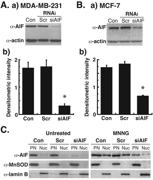
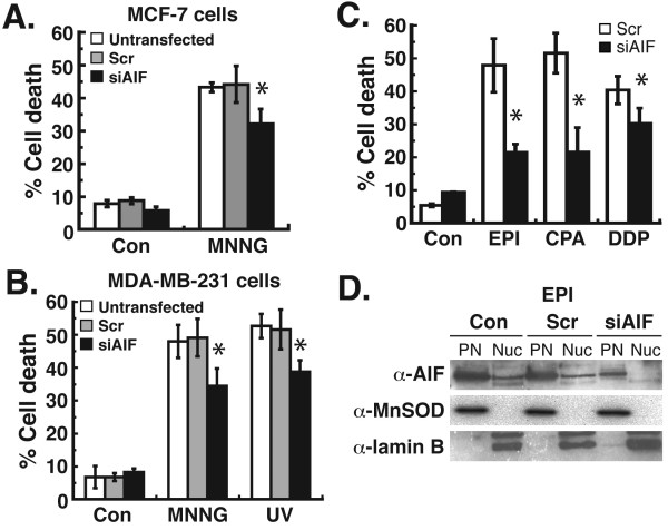
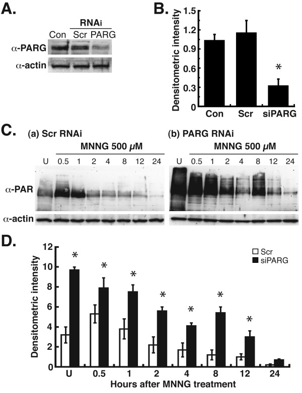
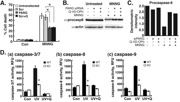
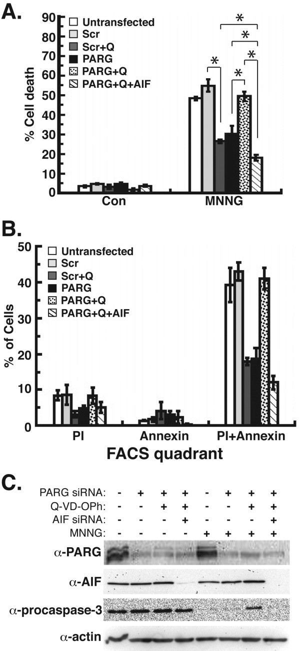
Results: Silencing of AIF by RNAi led to decreased MDA-MB-231 and MCF-7 breast cancer cell death after chemotherapy, which demonstrates a critical role for AIF. RNAi silencing of PAR glycohydrolase (PARG), the primary enzyme that catalyzes the hydrolysis of PAR, led to increased PAR levels but decreased cell death. Further investigation into the possible role of PAR in apoptosis revealed decreased caspase-3/7/8/9 activity in PARG-null cells. Interestingly, the pharmacologic inhibition of caspase activity in PARG-silenced breast cancer cells led to increased cell death after chemotherapy, which indicates that an alternative cell death pathway is activated due to elevated PAR levels and caspase inhibition. AIF silencing in these cells led to profound protection from chemotherapy, which demonstrates that the increased cell death after PARG silencing and caspase inhibition was mediated by AIF.

Conclusions: The results show a role for AIF in breast cancer cell death after chemotherapy, the ability of PAR to regulate caspase activity, and the ability of AIF to substitute as a primary mediator of breast cancer cell death in the absence of caspases. Thus, the induction of cell death by PAR/AIF may represent a novel strategy to optimize the eradication of breast tumors by activating an alternative cell death pathway.


Figures

Figure 1
Figure 1
Silencing of apoptosis-inducing factor (AIF) by siRNA in human breast adenocarcinoma cells. Human breast adenocarcinoma cells were transfected with 20 nM small interfering RNA (siRNA) oligos for AIF as previously reported [26]. A, (a) Immunoblotting detection of AIF in MDA-MB-231 cell extracts 48 h after siRNA transfection using polyclonal anti-AIF antibody (Rockland Immunochemcials) (1:1000 dilution). Controls were provided by untransfected cells (Con) and cells transfected with scrambled siRNA oligos (Scr). Densitometric quantification (b) of AIF protein bands from (a) was then performed. Values represent AIF/actin ratios of the densitometric intensities of the protein bands. *P < 0.01 between Scr and AIF (one-way ANOVA and unpaired Student’s t test). Error bars represent the standard error of the mean (SEM). B, Immunoblotting detection (a) and densitometric quantification of AIF in MCF-7 cell extracts 48 h after siRNA transfection. C, MDA-MB-231 cells were transfected with siRNA oligos, treated with 0.5 mM N-methyl-N’-nitro-N-nitrosoguanidine (MNNG) for 45 min, then analyzed by immunoblot for levels of nuclear AIF. Controls for cell fractionations were provided by mitochondrial manganese superoxide dismutase (MnSOD) and nuclear lamin B2. PN, post-nuclear fraction (cytoplasm + mitochondria); Nuc, nuclear fraction. All experiments were repeated at least three times with similar results.
Figure 2
Figure 2
Chemotherapeutic treatment of human breast adenocarcinoma cells after the silencing of AIF. Cells were transfected with siRNA for 48 h as before. A, MCF-7 cells were then treated with 0.5 mM MNNG x45 min. B, MDA-MB-231 cells were then treated with MNNG or 25 J/m2 UV-C radiation. After 24 h, cell death was quantified by FACS after propidium iodide (PI)/annexin V-FITC staining. C, MDA-MB-231 cells were treated with 20 μM epirubicin (EPI) x6 h, 10 mM cyclophosphamide (CPA) x24 h, or 0.5 mM cisplatin (DDP) x6 h after siRNA transfection. Cell death was then quantified by FACS after PI/annexin staining. For A-C, *P < 0.01 between Scr and AIF (one-way ANOVA and unpaired Student’s t test). Error bars represent the SEM. D, Analysis of nuclear AIF levels in siRNA transfected cells treated with EPI. Con, untransfected cells; Scr, scrambled siRNA transfected cells. All experiments were repeated at least twice with similar results.
Figure 3
Figure 3
Silencing of poly(ADP-ribose) glycohydrolase (PARG) by siRNA in MDA-MB-231 cells. MDA-MB-231 cells were transfected with siRNA oligos for PARG as previously reported [38]. A, Immunoblotting detection of PARG in cell extracts 48 h after transfection using polyclonal anti-PARG antibody (Millipore) (1:2,000 dilution). Equivalent controls wereutilized as in Fig. 1A. B, Densitometric quantification of PARG protein bands from A. *P < 0.01 between Scr and AIF (one-way ANOVA and unpaired Student’s t test). Error bars represent the SEM. C, MDA-MB-231 cells were transfected with PARG siRNA oligos, treated with 0.5 mM MNNG, then analyzed for levels of PAR by immunoblot from 0.5-24 h. Equal protein loading per lane was verified by immunoblotting detection of β-actin. D, Densitometric quantification of PAR levels from C. *P < 0.01 between Scr and PARG (one-way ANOVA and unpaired Student’s t-test). Error bars represent the SEM.
Figure 4
Figure 4
Analysis of cell death and caspases in MDA-MB-231 cells after PARG silencing or genetic disruption. A, MDA-MB-231 cells pretreated with the pan-caspase inhibitor, Q-VD-OPH (Q), were transfected with PARG siRNA. 48 h after transfection, cells were treated with 0.5 mM MNNG for 45 min, and 24 h later cell death was quantified by FACS. All error bars represent the SEM. *P < 0.01 between Scr and PARG or Scr and Scr + Q (one-way ANOVA and unpaired Student’s t-test). B, extracts from cells in A were probed for the immunoblotting detection of procaspase-8 levels. C, Procaspase-8 protein levels in B were quantified by densitometric analysis. Values represent procaspase-8/actin ratios of the densitometric intensities of the protein bands. D, Wild-type (WT) and PARG-null (KO) cells were treated with 25 J/m2 UV-C radiation and 12 h later caspase-3 (a), caspase-8 (b), and caspase-9 (c) activity was assayed. Q, pretreatment with Q-VD-OPh 30 min prior to UV treatment and until caspase activity assays. Error bars represent the SEM. *P < 0.01 between WT and KO (one-way ANOVA and unpaired Student’s t-test). RFU, relative fluorescence unit.
Figure 5
Figure 5
MDA-MB-231 cell death after silencing of PARG, inhibition of caspases, and RNAi knockdown of AIF. MDA-MB-231 cells were pretreated with Q-VD-OPh and transfected with siRNA for PARG and AIF. A, Cell death was quantified by FACS after treatment with 0.5 mM MNNG for 45 min. B, The percentage of cells that exhibited propidium iodide (PI), annexin V-FITC, or dual PI/annexin V-FITC fluorescence from FACS analysis in B were determined. C, Extracts from cells in A were probed for the immunoblotting detection of PARG, AIF, and procaspase-3. SDS-PAGE loading controls were provided by the immunoblotting detection of β-actin. All error bars represent the SEM. *P < 0.01 between Scr vs. Scr + Q, PARG vs. PARG + Q, PARG + Q vs. PARG + Q + AIF, PARG + Q + AIF vs. PARG, and PARG + Q + AIF vs. Scr + Q (one-way ANOVA and unpaired Student’s t-test).

References

    1. Juarez-Salinas H, Sims JL, Jacobson MK. Poly(ADP-ribose) levels in carcinogen-treated cells. Nature. 1979;282:740–741. doi: 10.1038/282740a0. - DOI - PubMed
    1. Miramar MD, Costantini P, Ravagnan L, Saraiva LM, Haouzi D, Brothers G, Penninger JM, Peleato ML, Kroemer G, Susin SA. NADH oxidase activity of mitochondrial apoptosis-inducing factor. J Biol Chem. 2001;276:16391–16398. doi: 10.1074/jbc.M010498200. - DOI - PubMed
    1. Pospisilik JA, Knauf C, Joza N, Benit P, Orthofer M, Cani PD, Ebersberger I, Nakashima T, Sarao R, Neely G. et al.Targeted deletion of AIF decreases mitochondrial oxidative phosphorylation and protects from obesity and diabetes. Cell. 2007;131:476–491. doi: 10.1016/j.cell.2007.08.047. - DOI - PubMed
    1. Joza N, Susin SA, Daugas E, Stanford WL, Cho SK, Li CY, Sasaki T, Elia AJ, Cheng HY, Ravagnan L. et al.Essential role of the mitochondrial apoptosis-inducing factor in programmed cell death. Nature. 2001;410:549–554. doi: 10.1038/35069004. - DOI - PubMed
    1. Artus C, Boujrad H, Bouharrour A, Brunelle MN, Hoos S, Yuste VJ, Lenormand P, Rousselle JC, Namane A, England P. et al.AIF promotes chromatinolysis and caspase-independent programmed necrosis by interacting with histone H2AX. EMBO J. 2010;29:1585–1599. doi: 10.1038/emboj.2010.43. - DOI - PMC - PubMed

Publication types

MeSH terms