. 2012 Nov;33(31):7785-93.
doi: 10.1016/j.biomaterials.2012.07.022. Epub 2012 Jul 26.

PET imaging of tumor associated macrophages using mannose coated 64Cu liposomes

Affiliations

PET imaging of tumor associated macrophages using mannose coated 64Cu liposomes

Landon W Locke et al. Biomaterials. 2012 Nov.

Abstract

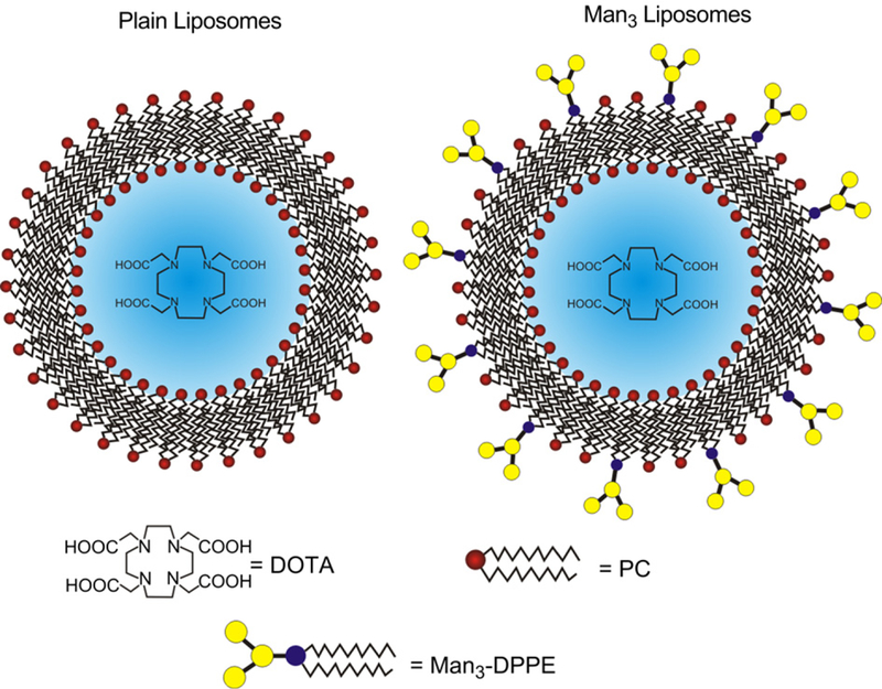
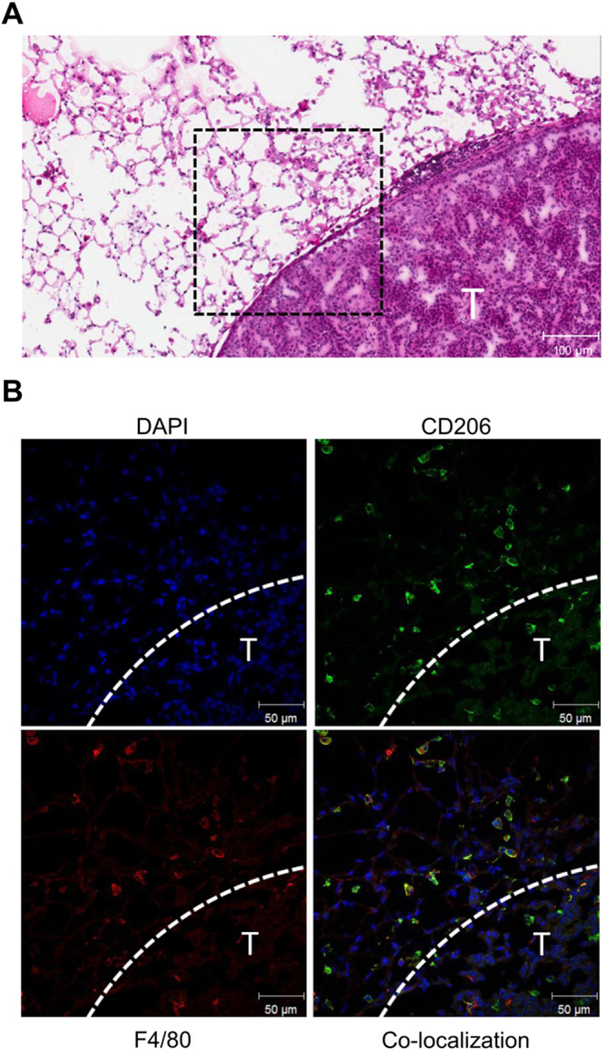
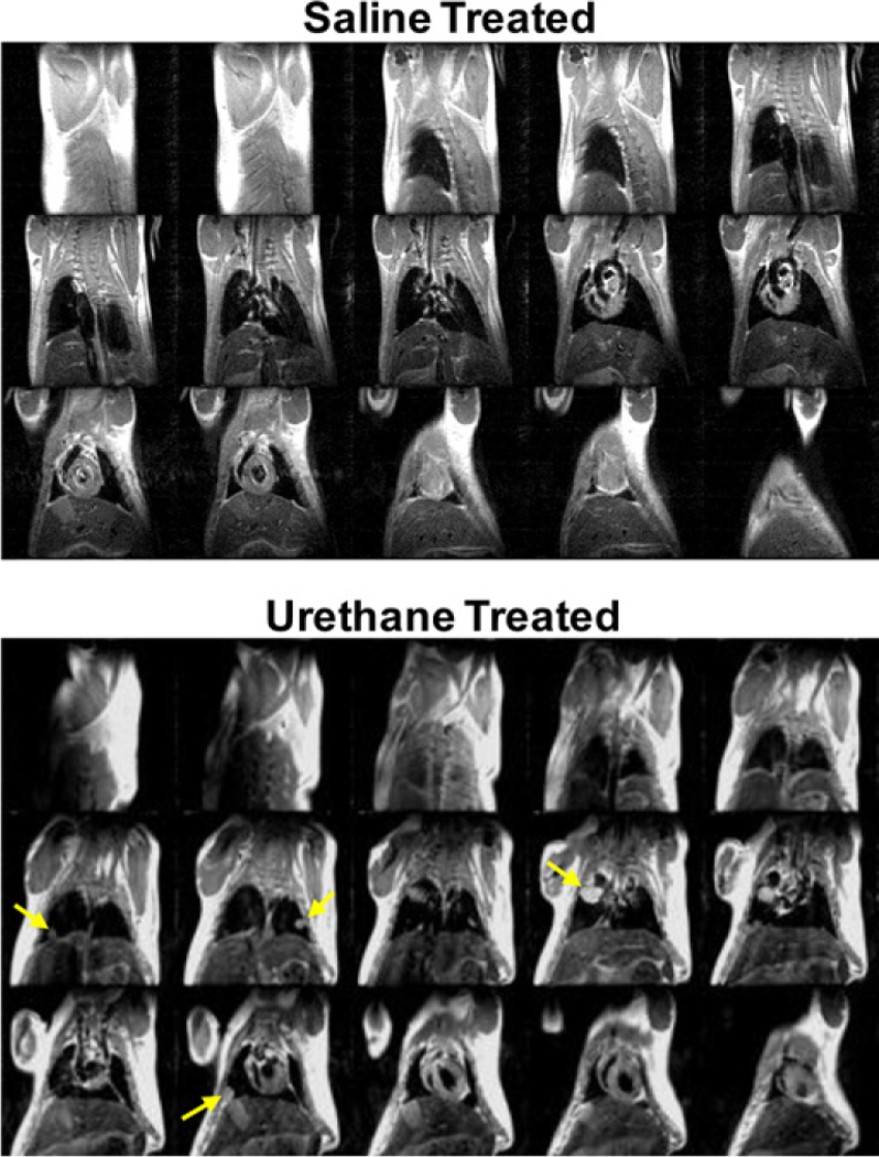
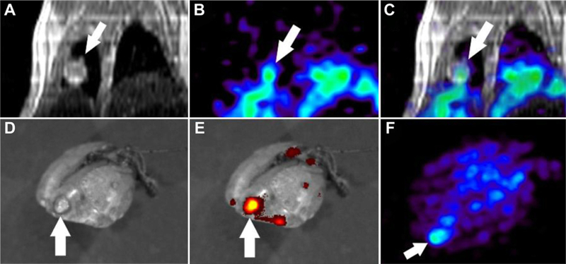
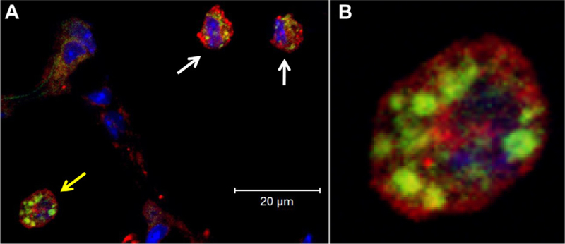
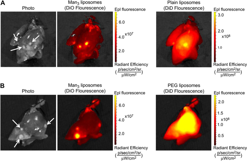
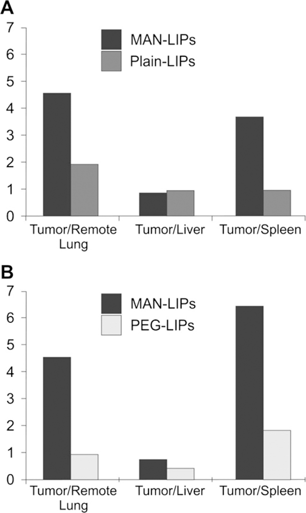
Macrophages within the tumor microenvironment (TAMs) have been shown to play a major role in the growth and spread of many types of cancer. Cancer cells produce cytokines that cause macrophages to express scavenger receptors (e.g. the mannose receptor) and factors that facilitate tissue and blood vessel growth, suppress T cell mediated anti-tumor activity, and express enzymes that can break down the extracellular matrix, thereby promoting metastasis. We have designed a mannosylated liposome (MAN-LIPs) and show that it accumulates in TAMs in a mouse model of pulmonary adenocarcinoma. These liposomes are loaded with (64)Cu to allow tracking by PET imaging, and contain a fluorescent dye in the lipid bilayer permitting subsequent fluorescence microscopy. We injected these liposomes into a mouse model of lung cancer. In vivo PET images were acquired 6 h after injection followed by the imaging of select excised organs. MAN-LIPs accumulated in TAMs and exhibited little accumulation in remote lung areas. MAN-LIPs are a promising new vehicle for the delivery of imaging agents to lung TAMs. In addition to imaging, MAN-LIPs hold the potential for delivery of therapeutic agents to the tumor microenvironment.


Figures

Fig. 1
Fig. 1
A schematic diagram of liposomes. DOTA-containing plain (left) and Man3 (right) liposomes allow for remote loading of the PET imaging agent, 64Cu. The mean liposome diameter was 200 nm.
Fig. 2
Fig. 2
Macrophage M2 polarization in lungs of a urethane-treated mouse 25 weeks after treatment. The H&E image shows a tumor boundary (T) with moderate immune cell infiltration in the stroma (A). Immunofluorescence staining of the tumor stroma outlined in the H&E image by the black dashed box is shown in (B). At the edge of the tumor (indicated by dashed white line), the majority of macrophages identified by F4/80 (red) also stain for CD206 (green) indicating M2 polarization.(For interpretation of the references to colour in this figure legend,the reader is referred to the web version of this article.)
Fig. 3
Fig. 3
Representative spin echo MR images of a saline (top) and urethane-injected (bottom) mouse 24 weeks following treatment. These scans were acquired approximately 30 min after gadolinium injection. Lungs tumors, as indicated by yellow arrows, are clearly visible in the urethane-treated mouse. In contrast, no lung tumors are detected in the saline-treated mouse. (For interpretation of the references to colour in this figure legend, the reader is referred to the web version of this article.)
Fig. 4
Fig. 4
Representative in vivo images showing a lung tumor on coronal MRI (A) with enhanced 64Cu-labeled Man3-liposome uptake on PET 6 h after i.v. injection (B). PET-MR image registration (C) verifies tumor localization of the PET signal. Ex vivo fluorescence image of the lung was obtained to assess DiO distribution. A photo shows the tumor (D) which exhibited higher DiO accumulation compared to non-tumor lung areas. Ex vivo PET maximum intensity projection image (F) showed focal 64Cu signal in the area of the lung spatially corresponding to the tumor shown in the photograph.
Fig. 5
Fig. 5
Confocal fluorescence microscopy revealed internalization of DiO-labeled Man3-liposomes (green) by F4/80+ macrophages (red) within the tumor stroma 6 h after i.v. liposome injection. Cell nuclei are stained with DAPI (blue). (A) A co-localized confocal image shows the intracellular localization of Man3-liposomes within TAMs. The enlarged view of the cell indicated by the yellow arrow is shown in (B), clearly shows a clustered distribution of liposomes consistent with storage in macrophage lysosomal structures. (For interpretation of the references to colour in this figure legend,the reader is referred to the web version of this article.)
Fig. 6
Fig. 6
Representative photos and fluorescent images from the liposome co-injection study. Images of excised lungs 6 h after the co-injection of Man3 and plain liposomes (A) and Man3 and PEG liposomes (B). Strong fluorescence signal associated with Man3 and plain liposomes is localized to lung tumors (identified by white arrows on the photo). However, compared to Man3-liposomes, plain liposomes exhibit a higher background signal. PEG liposomes show a diffuse lung distribution and consequently poor tumor contrast likely due to their enhanced blood circulation time.
Fig. 7
Fig. 7
Tumor-to-tissue ratios measured from fluorescence images of harvested organs following the co-injection of MAN and plain liposomes (A) and MAN and PEG lipo-somes (B). MAN-LIPs exhibited a higher tumor-to-remote lung and tumor-to-spleen ratio compared to plain liposomes, while tumor-to-liver ratios were comparable. MAN-liposomes also exhibited a high tumor-to-remote lung ratio following co-injection with PEG liposomes, likely due to the slow rate of blood clearance of PEG liposomes. PEG liposomes also showed a reduced tumor-to-liver ratio, consistent with a lower rate of capture by the RES.

References

    1. Marin L, Colombo P, Bebawy M, Young PM, Traini D, Marin L, et al. Chronic obstructive pulmonary disease: patho-physiology, current methods of treatment and the potential for simvastatin in disease management. Expert Opin Drug Deliv 2011;8(9):1205–20. - PubMed
    1. Murphy BS, Bush HM, Sun dareshan V, Davis C, Hagadone J, Cory TJ, et al. Characterization of macrophage activation states in patients with cystic fibrosis. J Cyst Fibros 2010;9(5):314–22. - PubMed
    1. Allavena P, Sica A, Solinas G, Porta C, Mantovani A, Allavena P, et al. The inflammatory micro-environment in tumor progression: the role of tumor-associated macrophages. Crit Rev Oncol Hematol 2008;66(1):1–9. - PubMed
    1. Bingle L, Brown NJ, Lewis CE, Bingle L, Brown NJ, Lewis CE. The role of tumour-associated macrophages in tumour progression: implications for new anti-cancer therapies. J Pathol 2002;196(3):254–65. - PubMed
    1. Murdoch C, Muthana M, Coffelt SB, Lewis CE, Murdoch C, Muthana M, et al. The role of myeloid cells in the promotion of tumour angiogenesis. Nat Rev Cancer 2008;8(8):618–31. - PubMed

Publication types