. 2012 Jul 26;2(1):89-100.
doi: 10.1016/j.celrep.2012.05.018. Epub 2012 Jul 5.

Differential trafficking of transport vesicles contributes to the localization of dendritic proteins

Affiliations

Differential trafficking of transport vesicles contributes to the localization of dendritic proteins

Sarmad Al-Bassam et al. Cell Rep. .

Abstract

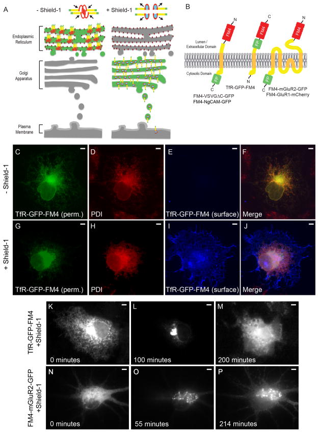
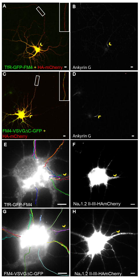
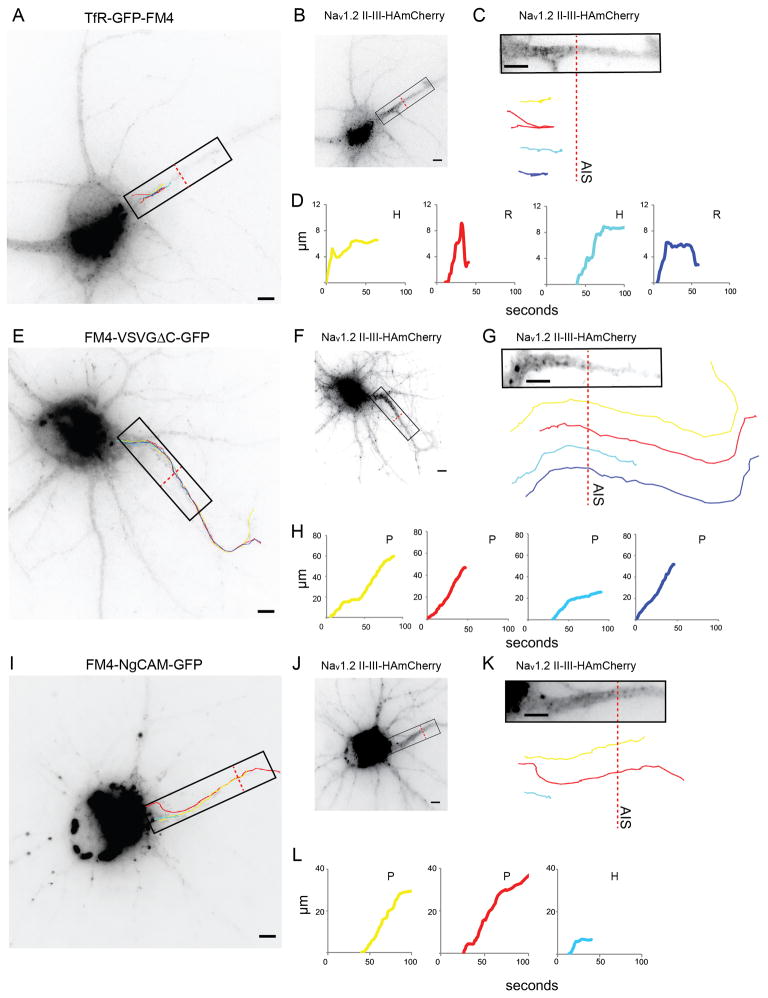
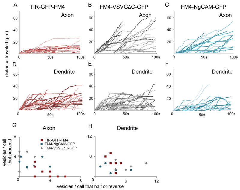
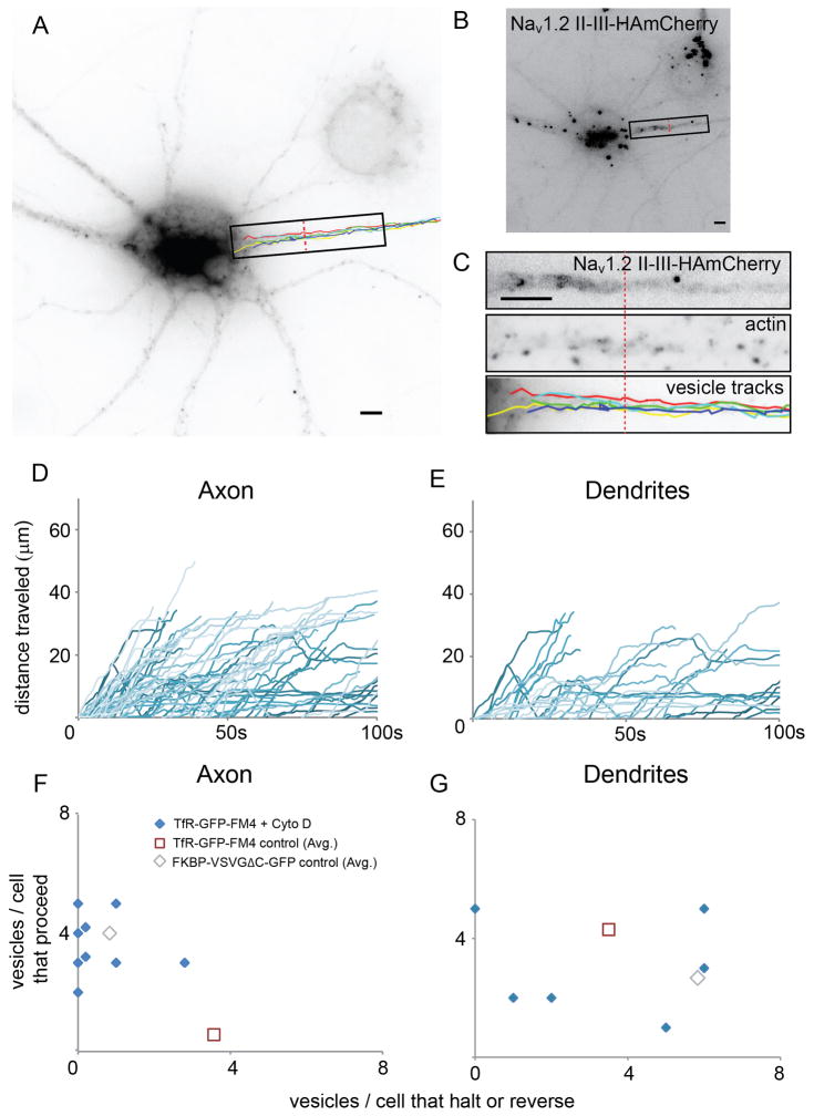
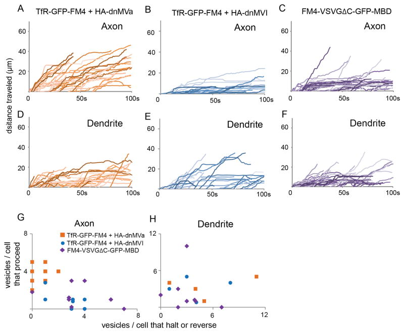
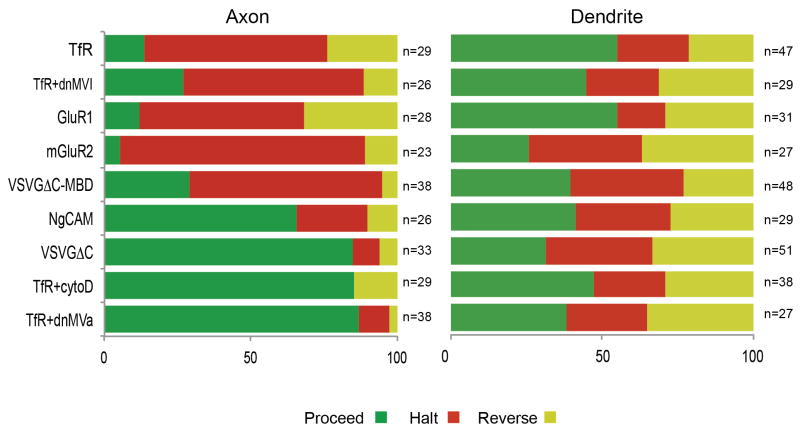
In neurons, transmembrane proteins are targeted to dendrites in vesicles that traffic solely within the somatodendritic compartment. How these vesicles are retained within the somatodendritic domain is unknown. Here, we use a novel pulse-chase system, which allows synchronous release of exogenous transmembrane proteins from the endoplasmic reticulum to follow movements of post-Golgi transport vesicles. Surprisingly, we found that post-Golgi vesicles carrying dendritic proteins were equally likely to enter axons and dendrites. However, once such vesicles entered the axon, they very rarely moved beyond the axon initial segment but instead either halted or reversed direction in an actin and Myosin Va-dependent manner. In contrast, vesicles carrying either an axonal or a nonspecifically localized protein only rarely halted or reversed and instead generally proceeded to the distal axon. Thus, our results are consistent with the axon initial segment behaving as a vesicle filter that mediates the differential trafficking of transport vesicles.


Figures

Figure 1
Figure 1
An FM4/Shield-1-based pulse chase system allows for synchronization of transmembrane proteins in the secretory pathway. (A) In the absence of Shield-1 transmembrane proteins (yellow) fused to FM4 domains (red) and to fluorescent proteins (green) multimerize causing clustering of the expressed fusion protein and retention within the ER. Binding of Shield-1 (cyan) to the FM4 domain causes disaggregation of the expressed transmembrane fusion protein, releasing it from the ER and allowing it to proceed through the secretory pathway. (B) Constructs contain 4 FM domains in tandem on the extracellular domain and a fluorescent protein fused to transmembrane proteins. (C, F) Following expression in COS cells and without exposure to Shield-1, TfR-GFP-FM4 (green) localizes in a reticular pattern that colocalizes with the endogenous ER markerPDI (red, D, F). A lack of TfR-GFP-FM4 on the surface (blue, E, F) is consistent with it being retained within the ER. In contrast, in cells exposed to Shield-1 TfR-GFP-FM4 (G, J) only partially colocalizes with PDI (H, J ) and TfR-GFP-FM4 expresses on the cell surface (I, J). Live imaging confirms that when TfR-GFP-FM4 is expressed in a COS cell for 12–16 hours it localizes in a reticular manner, suggesting that it is retained in the ER (K). Following addition of Shield-1 it becomes concentrated in a pattern consistent with Golgi localization (L). Subsequently, it localizes in a more diffuse pattern consistent with expression on the cell surface (M). Similarly, when FM4-mGluR2-GFP is expressed in a neuron for 16 hours, it localizes in a reticular manner consistent with retention in the ER (N). After the addition of Shield-1, FM4-mGluR2-GFP localizes in a perinuclear pattern, suggesting it had migrated to the Golgi (O). Afterwards, FM4-mGluR2-GFP shows a diffuse pattern of labeling consistent with presence on the plasma membrane (P). Scale bar is 5 μm.
Figure 2
Figure 2
Vesicles carrying TfR-GFP-FM4 enter axonal and dendritic processes with equal probability following exit from the Golgi (see also Figure S1, Movie 1). (A) In cortical neurons in dissociated culture that are exposed to Shield-1, TfR-GFP-FM4 (green) localizes in the somatodendritic compartment and is relatively absent from the axon as compared with HAmCherry (red), which localizes nonspecifically. Inset shows axonal region within the white box at higher magnification. (B) Ankyrin G staining labels the axonal process of the cell. (C) In contrast, both HAmCherry (red) and FM4-VSVGΔC-GFP (green) localize nonspecifically. Inset shows axonal region within the white box at higher magnification. (D) AnkyrinG staining labels the axonal process of the cell. (E) Tracks made by vesicles carrying TfR-GFP-FM4 following exit from the Golgi are consistent with these vesicles entering the closest process regardless of whether it is an axon or dendrite. (F) Nav1.2 II-III-HAmCherry co-expression labels the axonal process of the cell. (G) Vesicles containing FM4-VSVGΔC-GFP enter dendrites and axons with frequencies similar to that of vesicles carrying TfR-GFP-FM4 following exit from the Golgi. (H) Nav1.2 II-III-HAmCherry co-expression labels the axonal process of the cell. Arrowheads (yellow) point to the axon initial segment in each panel. Scale bar is 5 μm.
Figure 3
Figure 3
Vesicles carrying TfR-GFP-FM4 tend to haltand reverse within the AIS, whereas vesicles carrying FM4-VSVGΔC-GFP or FM4-NgCAM-GFPproceed beyond the end of the AIS (see also Figure S2 and Movies 2, 3). (A) Paths taken by vesicles carrying TfR-GFP-FM4 following release from the Golgi. (B) Localization pattern of Nav1.2 II-III-HAmCherry, which exhibits enrichment at the AIS. (C) Distribution of Nav1.2 II-III-HAmCherry in the AIS within the region shown in (A) compared with vesicle paths. (D) Graphs of distance traveled versus time for vesicle paths shown in (A) and (C). Note that all vesicles either halt or reverse before reaching the end of the AIS. (E) Paths taken by vesicles carrying FM4-VSVGΔC-GFP following release from the Golgi. (F) Localization pattern of Nav1.2 II-III-HAmCherry. (G) Distribution of Nav1.2 II-III-HAmCherry within the region shown in (E) compared with vesicle paths. (H) Graphs of distance versus time for vesicle paths shown in (E) and (G). Note that all vesicles proceed beyond the end of the AIS. (I) Paths taken by vesicles carrying FM4-NgCAM-GFP following release from the Golgi. ( J) Localization pattern of Nav1.2 II-III-HAmCherry. (K) Distribution of Nav1.2 II-III-HAmCherry within the region shown in (I).( L) Graphs of distance versus time for vesicle paths shown in (I) and(K ). Note that 2 vesicles proceed beyond the end of the AIS, while 1 vesicle halts. P refers to proceed, H to halt, R to reverse. Dashed red line marks the border of the AIS. Scale bar is 5 μm.
Figure 4
Figure 4
Vesicles carrying TfR-GFP-FM4 behave differently from those carrying FM4-VSVGΔC-GFP or FM4-NgCAM-GFP in axons, but similarly in dendrites (see also Figure S3 and Movies, 4, 5). (A) Distance traveled versus time plots for vesicles carrying TfR-GFP-FM4 in the axon reveal that most vesicles did not move more than 10 μm beyond the cell body following release from the Golgi. In dramatic contrast, similar plots for vesicles carrying FM4-VSVGΔC-GFP (B) reveal that these vesicles proceeded up to 60 μm into the axon. Similarly, plots for vesicles carrying FM4-NgCAM-GFP (C) reveal that these vesicles proceeded up to 50 μm into the axon. (D, E, F) Plots of distance traveled versus time for vesicles containing TfR-GFP-FM4 or FM4-VSVGΔC-GFP or FM4-NgCAM-GFP in dendrites indicate that all three types of vesicles behave in a similar manner in this compartment.( G, H) Axon and dendrite scatter plots showing the ratio of the number of vesicles that proceed versus the number that halt or reverse for individual cells. Note that each data point refers to a single cell. Vesicles carrying TfR-GFP-FM4 behave differently from those carrying FM-VSVGΔC-GFP or FM4-NgCAM-GFP in axons, but similarly in dendrites.
Figure 5
Figure 5
Actin filaments are necessary for halting and reversing of vesicles carrying TfR-GFP-FM4 in the AIS (see also Figure S4 and Movie 6). (A) Paths taken by vesicles carrying TfR-GFP-FM4 in the axons of cortical neurons in culture that were exposed to 4 μM cytochalasin D for 80 minutes. (B) AIS is defined by staining of Nav1.2 II-III-HAmCherry. (C) High power view of boxed region from (A) showing Nav1.2 II-III-HAmCherry, Phalloidin staining in AIS consistent with disrupted actin filaments, and vesicle tracks. (D) Plots of distance traveled versus time for vesicles in axons containing TfR-GFP-FM4 in cells exposed to cytochalasin D are consistent with those vesicles moving beyond the AIS into the distal axon. (E) Plots of distance versus time for vesicles in dendrites containing TfR-GFP-FM4 in cells exposed to cytochalasin D are similar to comparable plots of similar vesicles in control cells. (F, G) Axon and dendrite scatter plots showing the ratio of the number of vesicles carrying TfR-GFP-FM4 that proceed versus the number that halt or reverse for cells exposed to cytochalasin D. Note that each data point refers to a single cell. In axons such vesicles behave differently from similar vesicles in control cells, but similarly to those carrying FM4-VSVGΔC-GFP. In dendrites, vesicles carrying TfR-GFP-FM4 in cells exposed to cytochalasin D do not behave in a noticeably different manner from similar vesicles in control cells. Scale bar is 5 μm.
Figure 6
Figure 6
Interaction with Myosin Va, but not Myosin VI, is necessary for vesicle halting and reversing, whereas interaction with Myosin Va is sufficient to cause halting of vesicles at the AIS. (see also Figure S5 and Movies 7, 8). (A) Plots of distance traveled versus time for vesicles in axons containing TfR-GFP-FM4 in cells co-expressing HA-dnMVa are consistent with those vesicles moving beyond the AIS into the distal axon. In contrast, similar plots for vesicles in the axon containing TfR-GFP-FM4 in cells coexpressing HA-dnMVI (B) indicate that these vesicles are likely to halt or reverse while in the AIS. Note that this pattern is similar to that of cells expressing TfR-GFP-FM4 only. Similarly, plots of distance traveled versus time for vesicles in axons carrying FM4-VSVGΔC-GFP-MBD (C) indicate that those vesicles are more likely to halt within the AIS than vesicles carrying FM4-VSVGΔC-GFP. Plots of distance versus time show that vesicles in dendrites containing TfR-GFP-FM4 behave similarly in cells co-expressing HA-dnMVa (D) or HA-dnMVI (E). In addition, both types of vesicles behave in a similar manner to vesicles carrying FM4-VSVGΔC-GFP-MBD (F) in dendrites of control cells. (G, H) Axon and dendrite scatter plots showing the ratio of the number of vesicles carrying TfR-GFP-FM4 that proceed versus the number that halt or reverse in cells co-expressing HA-dnMVa or HA-dnMVI and a similar ratio for vesicles carrying FM4-VSVGΔC-GFP-MBD in control cells. Note that each data point refers to a single cell. In axons the vesicles carrying TfR-GFP-FM4 in cells co-expressing HA-dnMVI and vesicles carrying FM4-VSVGDC-GFP-MBD in control cells behave similarly to each other, but differently from those carrying TfR-GFP-FM4 in cells co-expressing HA-dnMVa. All three types of vesicles behave similarly in the dendrites.
Figure 7
Figure 7
In axon and dendrites, the overall percentage of vesicles that proceeded (green), halted (red) or reversed (yellow) with different contents orunder different conditions (see also Figure S6).

Similar articles

Cited by

References

    1. Alami NH, Jung P, Brown A. Myosin Va increases the efficiency of neurofilament transport by decreasing the duration of long-term pauses. J Neurosci. 2009;29:6625–6634. - PMC - PubMed
    1. Banaszynski LA, Chen LC, Maynard-Smith LA, Ooi AG, Wandless TJ. A rapid, reversible, and tunable method to regulate protein function in living cells using synthetic small molecules. Cell. 2006;126:995–1004. - PMC - PubMed
    1. Bridgman PC. Myosin Va movements in normal and dilute-lethal axons provide support for a dual filament motor complex. J Cell Biol. 1999;146:1045–1060. - PMC - PubMed
    1. Burack MA, Silverman MA, Banker G. The role of selective transport in neuronal protein sorting. Neuron. 2000;26:465–472. - PubMed
    1. Cai D, McEwen DP, Martens JR, Meyhofer E, Verhey KJ. Single molecule imaging reveals differences in microtubule track selection between Kinesin motors. PLoS Biol. 2009;7:e1000216. - PMC - PubMed

Publication types

LinkOut - more resources