

In vivo patellar tracking and patellofemoral cartilage contacts during dynamic stair ascending
- PMID: 22840488
- PMCID: PMC3438281
- DOI: 10.1016/j.jbiomech.2012.06.034
In vivo patellar tracking and patellofemoral cartilage contacts during dynamic stair ascending
Abstract
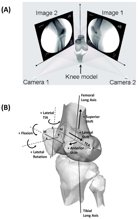
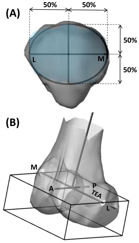
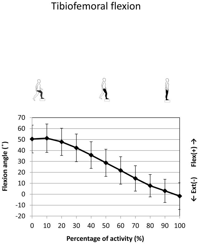
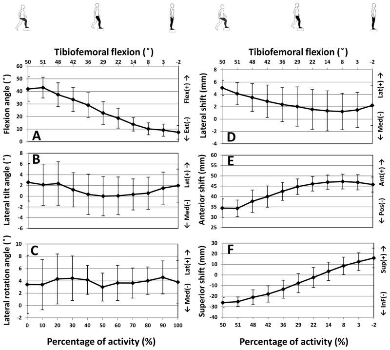
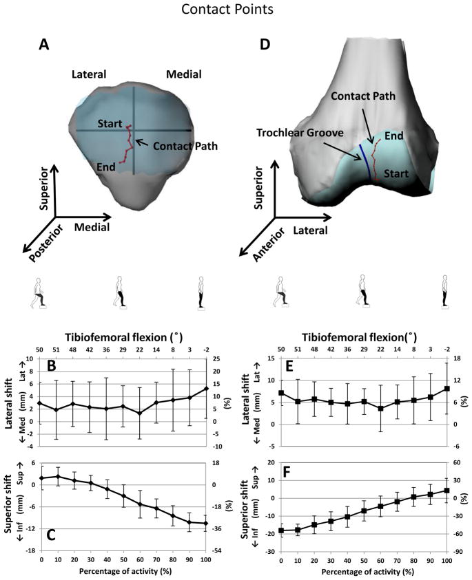
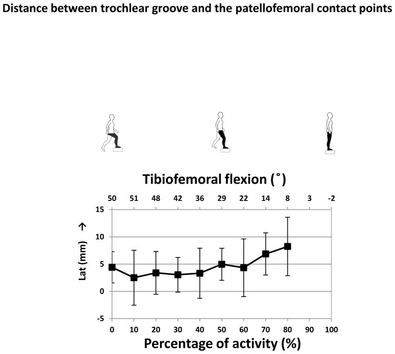
The knowledge of normal patellar tracking is essential for understanding the knee joint function and for diagnosis of patellar instabilities. This paper investigated the patellar tracking and patellofemoral joint contact locations during a stair ascending activity using a validated dual-fluoroscopic imaging system. The results showed that the patellar flexion angle decreased from 41.9° to 7.5° with knee extension during stair ascending. During first 80% of the activity, the patella shifted medially about 3.9 mm and then slightly shifted laterally during the last 20% of the ascending activity. Anterior translation of 13 mm of the patella was measured at the early 80% of the activity and the patella slightly moved posteriorly by about 2mm at the last 20% of the activity. The path of cartilage contact points was slightly lateral on the cartilage surfaces of patella and femur. On the patellar cartilage surface, the cartilage contact locations were about 2mm laterally from heel strike to 60% of the stair ascending activity and moved laterally and reached 5.3mm at full extension. However, the cartilage contact locations were relatively constant on the femoral cartilage surface (∼5mm lateral). The patellar tracking pattern was consistent with the patellofemoral cartilage contact location pattern. These data could provide baseline knowledge for understanding of normal physiology of the patellofemoral joint and can be used as a reference for clinical evaluation of patellofemoral disorders.
Copyright © 2012 Elsevier Ltd. All rights reserved.
Conflict of interest statement
No potential conflict of interest is declared.
Figures
Similar articles
-
Subject-specific evaluation of patellofemoral joint biomechanics during functional activity.Med Eng Phys. 2014 Sep;36(9):1122-33. doi: 10.1016/j.medengphy.2014.06.009. Epub 2014 Jul 3. Med Eng Phys. 2014. PMID: 24998901
-
The coupled effects of crouch gait and patella alta on tibiofemoral and patellofemoral cartilage loading in children.Gait Posture. 2018 Feb;60:181-187. doi: 10.1016/j.gaitpost.2017.12.005. Epub 2017 Dec 5. Gait Posture. 2018. PMID: 29248848 Free PMC article.
-
Stair climbing results in more challenging patellofemoral contact mechanics and kinematics than walking at early knee flexion under physiological-like quadriceps loading.J Biomech. 2009 Nov 13;42(15):2590-6. doi: 10.1016/j.jbiomech.2009.07.007. Epub 2009 Aug 4. J Biomech. 2009. PMID: 19656517
-
Research Methods and Progress of Patellofemoral Joint Kinematics: A Review.J Healthc Eng. 2019 Mar 24;2019:9159267. doi: 10.1155/2019/9159267. eCollection 2019. J Healthc Eng. 2019. PMID: 31019669 Free PMC article. Review.
-
Development of an innovative measurement method for patellar tracking disorder.Aging (Albany NY). 2020 Dec 1;13(1):516-524. doi: 10.18632/aging.202161. Epub 2020 Dec 1. Aging (Albany NY). 2020. PMID: 33260153 Free PMC article. Review.
Cited by
-
Preoperative flexion contracture is a predisposing factor for cartilage degeneration at the patellofemoral joint after open wedge high tibial osteotomy.Knee Surg Relat Res. 2020 Oct 13;32(1):55. doi: 10.1186/s43019-020-00063-2. Knee Surg Relat Res. 2020. PMID: 33050942 Free PMC article.
-
HKA angle exceeding 5 degrees is strongly associated with lateral patellar translation beyond 2 mm: surgical recommendations for avoiding adverse effects on the patellofemoral joint after OWHTO.J Orthop Surg Res. 2025 Jan 15;20(1):45. doi: 10.1186/s13018-024-05391-7. J Orthop Surg Res. 2025. PMID: 39810245 Free PMC article.
-
In vivo patellofemoral contact mechanics during active extension using a novel dynamic MRI-based methodology.Osteoarthritis Cartilage. 2013 Dec;21(12):1886-1894. doi: 10.1016/j.joca.2013.08.023. Epub 2013 Sep 3. Osteoarthritis Cartilage. 2013. PMID: 24012620 Free PMC article.
-
Patellofemoral Mechanics: a Review of Pathomechanics and Research Approaches.Curr Rev Musculoskelet Med. 2020 Jun;13(3):326-337. doi: 10.1007/s12178-020-09626-y. Curr Rev Musculoskelet Med. 2020. PMID: 32394363 Free PMC article. Review.
-
Impact of five different medial patellofemoral ligament-reconstruction strategies and three different graft pre-tensioning states on the mean patellofemoral contact pressure: a biomechanical study on human cadaver knees.J Exp Orthop. 2018 Jun 28;5(1):25. doi: 10.1186/s40634-018-0140-x. J Exp Orthop. 2018. PMID: 29956015 Free PMC article.
References
-
- Ahmed AM, Duncan NA, Tanzer M. In Vitro Measurement of the Tracking Pattern of the Human Patella. J Biomech Eng. 1999;121(2):222–228. - PubMed
-
- Amis AA, Senavongse W, Bull AM. Patellofemoral Kinematics During Knee Flexion-Extension: An in Vitro Study. J Orthop Res. 2006;24(12):2201–2211. - PubMed
-
- Beck PR, Thomas AL, Farr J, Lewis PB, Cole BJ. Trochlear Contact Pressures after Anteromedialization of the Tibial Tubercle. Am J Sports Med. 2005;33(11):1710–1715. - PubMed
-
- Brossmann J, Muhle C, Schroder C, Melchert UH, Bull CC, Spielmann RP, Heller M. Patellar Tracking Patterns During Active and Passive Knee Extension: Evaluation with Motion-Triggered Cine Mr Imaging. Radiology. 1993;187(1):205–212. - PubMed
-
- Churchill DL, Incavo SJ, Johnson CC, Beynnon BD. The Transepicondylar Axis Approximates the Optimal Flexion Axis of the Knee. Clin Orthop Relat Res. 1998;(356):111–118. - PubMed
Publication types
MeSH terms
Grants and funding
LinkOut - more resources
Full Text Sources
