. 2012 Jul 27;19(7):819-30.
doi: 10.1016/j.chembiol.2012.05.022.

Arginine topology controls escape of minimally cationic proteins from early endosomes to the cytoplasm

Affiliations

Arginine topology controls escape of minimally cationic proteins from early endosomes to the cytoplasm

Jacob S Appelbaum et al. Chem Biol. .

Abstract

Proteins represent an expanding class of therapeutics, but their actions are limited primarily to extracellular targets because most peptidic molecules fail to enter cells. Here we identified two small proteins, miniature protein 5.3 and zinc finger module ZF5.3, that enter cells to reach the cytosol through rapid internalization and escape from Rab5+ endosomes. The trafficking pathway mapped for these molecules differs from that of Tat and Arg(8), which require transport beyond Rab5+ endosomes to gain cytosolic access. Our results suggest that the ability of 5.3 and ZF5.3 to escape from early endosomes is a unique feature and imply the existence of distinct signals, encodable within short sequences, that favor early versus late endosomal release. Identifying these signals and understanding their mechanistic basis will illustrate how cells control the movement of endocytic cargo and may allow researchers to engineer molecules to follow a desired delivery pathway for rapid cytosolic access.


Figures

Figure 1
Figure 1
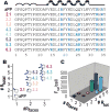
Miniature protein design. (A) Sequences of cationic miniature proteins evaluated in this work. (B) A plot of the relationship between #Rtotal (the number of (-helical arginine residues) and #Rfaces (the number of α-helical faces on which these arginines are displayed) for each aPP variant. The location of each α–helical arginine residue is represented by a blue circle on the helical wheel. (C) A plot of mean cellular fluorescence at 530 nm of HeLa cells treated with fluorescently tagged miniature protein variants (5 μM, 30 min). See also Figure S1.
Figure 2
Figure 2
Arginine topology controls cell binding and uptake. (A) Surface binding of rhodamine labeled cationic miniature proteins in the absence of endocytosis and removal by trypsin treatment. HeLa cells were treated with 1 μM rhodamine labeled cationic miniature protein for 30 min. Cell were then treated with trypsin (0.05%, 10 min, 37°C) or PBS before washing and analysis by flow cytometry. (B) Fraction of cell-associated fluorescence remaining after trypsin treatment. These data represent the ratio of black to red bars shown in A. (C) Cell uptake of rhodamine labeled peptides by HeLa cells after 30 min and 90 min.AlexaFluor-488-transferrin (Tf488) when added to HeLa cells (D) colocalizes with (E) Tf546 and (F) rhodamine labeled miniature proteins. Perfect colocalization is characterized by a Pearson's R value (R) equal to 1, while R values near 0 represent little or no colocalization. The correlation value observed when cells were treated with both Tf488 and alexa-fluor-546-labeled transferrin (Tf46) is 0.905. (G) When added with Tf488, aPPR shows little intracellular signal. Rhodamine labeled cationic miniature proteins and Tf546 are shown in red, Tf488 is shown in green, Hoescht (nucleus) is shown in blue. See also Figure S2.
Figure 3
Figure 3
Translocation of GR-GFP after treatment with dexamethasone and dexamethasone labeled peptides but not aPPDex. (A) HeLa cells transfected with GR-GFP (which appears black in the top row) after no treatment (−), or treatment with 1 μM dexamethasone or 1 μM aPPDex for 30 min at 37°C. The lower panel is an overlay of the GFP signal, shown in green, and the nuclear Hoescht signal shown in blue. (B) Quantification scheme. (C) Change in GR-GFP after treatment with 1 μM Dex-labeled cationic miniature protein for 30 min. (D) Quantification of the changes visible in (c). ns, not significant. * p ≤ 0.05, *** p ≤ 0.001, ANOVA. See also Figure S3.
Figure 4
Figure 4
Endocytosis is required for cytosolic access. (A) Endocytosis inhibitors block the uptake of 5.3R, TatR and Arg8R. Translocation of GR-GFP after treatment with 1 μM Dex, 5.3Dex, TatDex, Arg8Dex, or aPPDex in the presence (gray) or absence (black) of various small molecule inhibitors. Inhibitors of endocytosis included (B) 80 μM dynasore, (C) 5 mM methyl-β-cyclodextrin (MβCD), (D) 50 μM EIPA. (E) Translocation ratio after treatment with cationic miniature proteins and 200 nM bafilomycin . * p ≤ 0.05; *** p ≤ 0.001, ANOVA with Bonferroni post test. See also Figure S4.
Figure 5
Figure 5
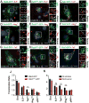
Miniature protein 5.3R enters via endocytosis into Rab5+ vesicles before trafficking to Rab7+ vesicles. HeLa cells transfected with the indicated GFP fusion protein (panels A-I) were treated with 1 μM 5.3R (panels A-C), TatR (panels D-F) or Arg8R (panels G-I) before being washed and imaged by confocal microscopy. Colocalization of 5.3R, TatR and Arg8R with Rab5-GFP is moderate (panels A, D, G) but can be increased by arresting Rab5 maturation via overexpression of Rab5Q79L-GFP. (panels B, E, H). 5.3R, TatR and Arg8R are delivered to Rab7+ endosomes (panels C, F, I). (J) Transfection with Rab5Q79L-GFP does not block the increase of translocation ratio seen after treatment with dexamethasone, 5.3Dex, or Arg8Dex, but decreases the translocation ratio measured after treatment with TatDex (p = 0.005). (K) HeLa cells treated with 200 nM wortmannin for 30 min before treatment with 1 μM Dex, 5.3Dex, TatDex, Arg8Dex, or aPPDex for 30 additional min in the continued presence of the drug. Wortmannin decreased the translocation ratio measured after treatment with TatDex (p = 8.1 ×10⁻¹⁵) and Arg8Dex (p = 2.3 ×10⁻⁵). ns, not significant; * p ≤ 0.05; ** p ≤ 0.01; *** p ≤ 0.001, ANOVA with Bonferroni post test. See also Figure S5.
Figure 6
Figure 6
The 5.3 Arginine Motif is Transportable. (A) Primary sequences of parent zinc finger (ZF), with charged residues mutated to alanine, and zinc finger displaying 5.3 arginine motif (ZF5.3). (B) Translocation of GR-GFP after treatment with 1 μM Dex-labeled zinc fingers in the presence and absence of endocytic inhibitors, as described in Figure 5. (C) Colocalization of Rab5-GFP with rhodamine labeled zinc fingers. Rhodamine labeled ZF5.3 shown in red, Rab5-GFP in green, Hoescht 33342 in blue. (D) Rab5Q79L-GFP overexpression does not block the increase of translocation ratio seen after treatment with ZF 5.3Dex. ns, not significant. *** p ≤ 0.001, ANOVA with Bonferroni post test. See also Figure S6.
Figure 7
Figure 7
Scheme illustrating the stepwise pathway of traffic taken by 5.3, ZF5.3, Tat and Arg8 from the cell exterior into the cytosol. Endocytosis from the plasma membrane is required for all three molecules to reach the cytoplasm. Endosomes rapidly acquire Rab5, followed by Vps34, which phosphorylates phosphoinositide lipids forming PI3P. Rab5 and PI3P recruit Rab5 effectors beginning endosome maturation. Recruitment of Rab7 leads to the formation of late endosomes (LE), but the Rab5+ to Rab7+ transition is not required for 5.3 or ZF5.3 to access the cytoplasm. Wortmannin treatment blocks Vps34 and recruitment of Rab5 effectors as well as Tat and Arg8 escape. Rab5Q79L acts later, allowing escape of Arg8 but not Tat. Endosomes are progressively acidified, a process blocked by Bafilomycin (structure shown) and required for 5.3, ZF5.3, Tat, and Arg8 to reach the cytoplasm.

References

    1. Androlewicz MJ, Anderson KS, Cresswell P. Evidence that transporters associated with antigen processing translocate a major histocompatibility complex class I-binding peptide into the endoplasmic reticulum in an ATP-dependent manner. Proc Natl Acad Sci USA. 1993;90:9130–9134. - PMC - PubMed
    1. Belitsky JM, Leslie SJ, Arora PS, Beerman TA, Dervan PB. Cellular uptake of N-methylpyrrole/N-methylimidazole polyamide-dye conjugates. Bioorg Med Chem. 2002;10:3313–3318. - PubMed
    1. Blundell TL, Pitts JE, Tickle IJ, Wood SP, Wu CW. X-ray analysis (1. 4-A resolution) of avian pancreatic polypeptide: Small globular protein hormone. Proc Natl Acad Sci USA. 1981;78:4175–4179. - PMC - PubMed
    1. Cantalupo G, Alifano P, Roberti V, Bruni CB, Bucci C. Rab-interacting lysosomal protein (RILP): the Rab7 effector required for transport to lysosomes. EMBO J. 2001;20:683–693. - PMC - PubMed
    1. Carey KL, Richards SA, Lounsbury KM, Macara IG. Evidence using a green fluorescent protein-glucocorticoid receptor chimera that the Ran/TC4 GTPase mediates an essential function independent of nuclear protein import. J Cell Biol. 1996;133:985–996. - PMC - PubMed

Publication types