. 2012 Jul 26;75(2):271-82.
doi: 10.1016/j.neuron.2012.05.022.

Photochemical restoration of visual responses in blind mice

Affiliations

Photochemical restoration of visual responses in blind mice

Aleksandra Polosukhina et al. Neuron. .

Abstract

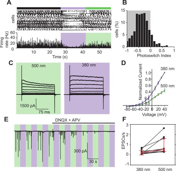
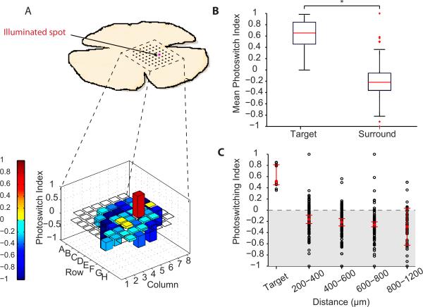
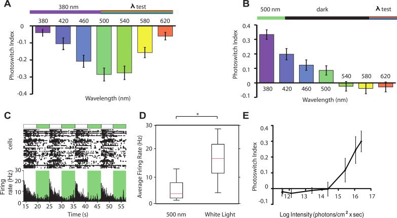
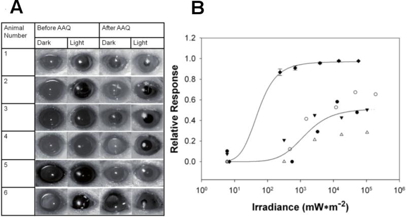
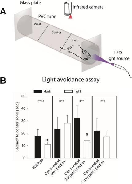
Retinitis pigmentosa (RP) and age-related macular degeneration (AMD) are degenerative blinding diseases caused by the death of rods and cones, leaving the remainder of the visual system intact but largely unable to respond to light. Here, we show that AAQ, a synthetic small molecule photoswitch, can restore light sensitivity to the retina and behavioral responses in vivo in mouse models of RP, without exogenous gene delivery. Brief application of AAQ bestows prolonged light sensitivity on multiple types of retinal neurons, resulting in synaptically amplified responses and center-surround antagonism in arrays of retinal ganglion cells (RGCs). Intraocular injection of AAQ restores the pupillary light reflex and locomotory light avoidance behavior in mice lacking retinal photoreceptors, indicating reconstitution of light signaling to brain circuits. AAQ and related photoswitch molecules present a potential drug strategy for restoring retinal function in degenerative blinding diseases.


Figures

Figure 1
Figure 1. AAQ imparts light sensitivity onto blind retinas from rd1 mice
(A) Multi-electrode recordings from flat-mounted rd1 mouse retinas before and after treatment with AAQ (300 μM for 25 min, followed by washout). Top, raster plot of spiking from RGCs; bottom, average RGC firing rate calculated in 100 msec time bins. Color bars represent illumination with 380 nm (violet) or 500 nm light (green), separated by periods of darkness. (B) Analysis of photoswitching of the entire population of RGCs from all untreated retinas and all AAQ-treated retinas. Untreated retinas (n=12) had PI values near 0, indicating no photoswitching, AAQ-treated retinas (n=21) had PI values >0, indicating an increase in firing frequency after switching from darkness to 380 nm light. (C) AAQ-mediated photosensitivity results from an increase in firing rate in 380 nm light. Average RGC firing rates in untreated retinas and AAQ-treated retinas in darkness and during the first 5 s in 380 nm light. Note that untreated retinas (n=12) fail to respond to light, but AAQ-treated retinas have RGCs that increase firing rate with 380 nm light. Red symbols show median values and error bars represent 95% confidence intervals for untreated and treated retinas (p < 0.0001, Mann-Whitney test).
Figure 2
Figure 2. Multiple types of retinal neurons contribute to the AAQ-mediated light response of RGCs
(A) Amacrine cell-mediated synaptic inhibition dominates the RGC light response. MEA recording with antagonists of GABAA (gabazine; 4 μM), GABAC (TPMPA; 10 μM), and glycine receptors (strychnine; 10 μM). Top, raster plot of RGC spiking. Bottom, average RGC firing rate. (B) After blocking inhibition, PI values show a decrease in firing frequency upon switching from darkness to 380 nm light (n=11 retinas). (C) Endogenous K+ channels contribute to the RGC light response. Whole-cell patch clamp recording from an RGC. Currents were evoked by voltage steps from -80 to +40 mV in 20-mV increments in 380 nm and 500 nm light. Inhibitory GABAergic and glycinergic inputs were blocked as in (a), and excitatory glutamatergic inputs were blocked with DNQX (10 μM) and AP5 (50 μM). (D). Photoregulation of endogenous K+ channels evaluated in steady-state I-V curves obtained in 380 and 500 nm light (n = 5 RGCs). Current is normalized to the maximal value at +40 mV (380 nm light). Variability among data is expressed as mean ± SEM. (E) Bipolar cell-mediated synaptic excitation also contributes to the RGC light response. Whole-cell patch clamp recording from an RGC. Blockade of inhibitory synaptic inputs (as in panel A) and endogenous RGC K+ channels (as in panel C) reveals photoregulation of EPSCs. Note the disappearance of EPSCs after superfusion with glutamate receptor antagonists DNQX (10 μM) and AP5 (50 μM). Holding potential = -60 mV. (F) Average EPSC rate in 380 nm and 500 nm light. Note the significant increase in EPSC rate in 500 nm light (p < 0.05, Mann-Whitney test; n=9 cells). Red symbols show median values and error bars represent 95% confidence intervals.
Figure 3
Figure 3. The AAQ treated retina generates spatially precise light responses
(A) Targeted illumination of a portion of the retinal centered on a single MEA electrode (top). The target (electrode E6) was exposed to 3 s flashes of alternating 380 and 500 nm light. ,Spot size = 60 μm in radius, inter-electrode spacing = 200 μm. Only the targeted electrode records an increase in RGC firing in response to 380 nm light (bottom). PI values are color-coded (scale at left) and also represented by bar height. Red bar is electrode E6 (PI = 0.812; n = 1 cell) and blue electrodes are the surround (PI = -0.209; n = 56 cells). Empty squares are electrodes on which no action potentials were recorded. (B) Targeted illumination results from 3 retinas, displayed in a box plot. PI values for the target and the surround RGCs are significantly different from one another (p < 0.005, Mann-Whitney test (C) Targeted illumination results in opposite responses in center and surround RGCs (n = 11 cells and n = 385 cells, respectively, from 3 retinas. PI values of RGCs (open circles) as a function of distance from the target electrode, displayed in 200 um bins. The red diamonds indicate the median plus or minus the bootstrapped 95% confidence intervals.
Figure 4
Figure 4. Spectral and illuminance sensitivity of AAQ-mediated photocontrol of RGC firing
(A) Spectral sensitivity of light-elicited suppression of RGC firing. Top: Light stimulation protocol. AAQ was first driven into its cis configuration with 380 nm light (5 s) and various test wavelengths triggered photoisomerization to the trans configuration. Bottom: PI values reveal the effectiveness of different wavelengths in suppressing RGC firing (n=5 retinas). (B) Spectral sensitivity of light-elicited activation of RGC firing. Top: Light stimulation protocol. AAQ was first driven into its trans configuration with 500 nm light (15 s). After an additional dark period (45 s) various test wavelengths triggered photoisomerization to the cis configuration. Bottom: PI values reveal the effectiveness of different wavelengths in stimulating RGC firing (n = 5 retinas). For (A) and (B) the PI was measured over the first 1 s after applying the test wavelength. (C) Stimulation of RGC firing in an AAQ-treated retina with white light. Top, raster plot of spiking from RGCs; bottom, average RGC firing rate. (D) Box plot representation of increased firing rate in white light vs. 500 nm. White light significantly increases peak firing rate (p < 0.05, Mann-Whitney test, n = 5). (E) Light intensity-response relationship for AAQ-treated rd1 mouse retinas exposed to different intensities of 380 nm light. Minimum light intensity needed for photoswitching is 2.6*1015 photons/cm2/sec.
Figure 5
Figure 5. AAQ restores the pupillary light response in mice lacking all retinal photoreceptors
(A) Pupillary light responses to 5.5 * 104 mW/m2 white light in opn4-/- rd/rd mice, before (left) and 3 hours after (right) intravitreal injection of AAQ (1 μl of 80 mM in DMSO). Dark images taken 5 s before light stimulus; light images represent maximal pupillary constriction during 30 s light exposure. Images were taken with an infrared-sensitive camera under infrared illumination. (B) Irradiance-dependence of pupillary light responses to white light. Irradiance response for wild-type mice (plotted as mean ± STD, n = 5) (◆) and four opn4-/- rd/rd mice injected with AAQ (plotted individually: ●○▼△). Data were fitted with a three parameter Hill equation.
Figure 6
Figure 6. AAQ restores active light avoidance behavior in mice lacking all retinal photoreceptors
(A) Schematic diagram of the locomotory light-avoidance test chamber. (B) Restoration of light avoidance behavior in opn4-/- rd/rd mice following AAQ injection. Bars represent mean latency of movement from the “East” to the “Center” third of the tube (plotted as ± STD).
Figure 7
Figure 7. AAQ restores light-modulated locomotor behavior in an open-field test
(A) Paths traveled by an rd1 mouse before and after injection with AAQ in darkness and with 380 nm illumination. (B) Cumulative distance traveled by the mouse in darkness and in 380 nm light, before and after AAQ. (C) Average cumulative distance traveled of all mice in darkness and 380 nm light, before and after AAQ. Closed squares represent time spent in darkness while open squares represent time spent in 380 nm light. (mean ± SEM, n = 8 ). (D) Mean locomotory velocity in light normalized to basal velocity in darkness. Velocity decreases significantly in light (n = 8, p < 0.0006). (E) Light evoked change in the velocity of each of the eight mice, before and after AAQ. Red line shows the mean light evoked change, before and after AAQ. (F) Light induced behavior is correlated with the light induced change in firing rate. Data were from the five mice for which both in vivo behavioral measurement and ex vivo retinal MEA recordings were obtained (as labeled in panel (E)). The light induced percent change in firing rate was calculated from the aggregate light response for all units recorded with the MEA upon switching from darkness to 380 nm light,. The light induced behavior represents percent change in velocity upon switching from darkness to 380 nm light.

Comment in

References

    1. Ahuja AK, Dorn JD, Caspi A, McMahon MJ, Dagnelie G, Dacruz L, Stanga P, Humayun MS, Greenberg RJ. Blind subjects implanted with the Argus II retinal prosthesis are able to improve performance in a spatial-motor task. Br J Ophthalmol. 2011;95:539–543. - PMC - PubMed
    1. Banghart MR, Mourot A, Fortin DL, Yao JZ, Kramer RH, Trauner D. Photochromic blockers of voltage-gated potassium channels. Angew Chem Int Ed Engl. 2009;48:9097–9101. - PMC - PubMed
    1. Beltran WA, Boye SL, Boye SE, Chiodo VA, Lewin AS, Hauswirth WW, Aguirre GD. rAAV2/5 gene-targeting to rods:dose-dependent efficiency and complications associated with different promoters. Gene Ther. 2010;17:1162–1174. - PMC - PubMed
    1. Benav H, Bartz-Schmidt KU, Besch D, Bruckmann A, Gekeler F, Greppmaier U, Harscher A, Kibbel S, Kusnyerik A, Peters T, et al. Restoration of useful vision up to letter recognition capabilities using subretinal microphotodiodes.. Engineering in Medicine and Biology Society (EMBC), 2010 Annual International Conference of the IEEE.2010. pp. 5919–5922. - PubMed
    1. Bi A, Cui J, Ma YP, Olshevskaya E, Pu M, Dizhoor AM, Pan ZH. Ectopic expression of a microbial-type rhodopsin restores visual responses in mice with photoreceptor degeneration. Neuron. 2006;50:23–33. - PMC - PubMed

Publication types

MeSH terms