. 2012 Jul 26;75(2):283-93.
doi: 10.1016/j.neuron.2012.05.019.

Restoration of hearing in the VGLUT3 knockout mouse using virally mediated gene therapy

Affiliations

Restoration of hearing in the VGLUT3 knockout mouse using virally mediated gene therapy

Omar Akil et al. Neuron. .

Abstract

Mice lacking the vesicular glutamate transporter-3 (VGLUT3) are congenitally deaf due to loss of glutamate release at the inner hair cell afferent synapse. Cochlear delivery of VGLUT3 using adeno-associated virus type 1 (AAV1) leads to transgene expression in only inner hair cells (IHCs), despite broader viral uptake. Within 2 weeks of AAV1-VGLUT3 delivery, auditory brainstem response (ABR) thresholds normalize, along with partial rescue of the startle response. Lastly, we demonstrate partial reversal of the morphologic changes seen within the afferent IHC ribbon synapse. These findings represent a successful restoration of hearing by gene replacement in mice, which is a significant advance toward gene therapy of human deafness.


Figures

Figure 1
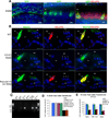
Figure 1. AAV1-GFP transduction in mice organ of Corti
1A. AAV1-GFP was used to assess viral delivery to the cochlea on organ of Corti surface preparations. AAV1 transfects a number of cell types including inner (IHC) and supporting cells (white arrows, left panel) in the organ of Corti. The VGLUT3 KO (V3KO, 1A middle panel) documents IHC labeling of transfected cells only. In the WT mouse (1A right panel), VGLUT3 co-labels IHCs (red) along AAV1- GFP transfected cells (green). 1B. VGLUT3 expression following AAV1-VGLUT3 delivery via the RWM, delivered at P10-12, and stained with anti-myosin 7A antibody (green, used as a hair cell marker, left column), anti-VGLUT3 antibody (red, middle column) and merged (right column). WT mice show IHCs labeled with both anti-Myo7A and VGLUT3 as expected (1B upper right panel) whereas KO mice only show Myo7a expression (1B middle right panel). Following AAV1-VGLUT3 delivery, the IHC is co-labeled by both anti-Myo7a and anti-VGLUT3 antibodies (1B lower right panel) (TC=tunnel of Corti, DC=Deiter‘s cells, OHC=outer hair cells, IHC=inner hair cells). 1C. RT-PCR was used to verify VGLUT3 mRNA expression in the transfected, KO (first lane) and WT (2nd lane) mice. Rescued whole cochlear extract (R-Co) demonstrates strong VGLUT3 mRNA expression, as does the stria vascularis + organ of Corti lane (R-SV+OC) and vestibular neuroepithelium (R-Vest). Spiral ganglion only shows weak mRNA expression (R-SG). 1D-E. Inner hair cells labeled with anti-VGLUT3 antibody following transfection were counted, tabulating both the entire cochlea and within the base, mid-turn and apex to determine differences in regions (at P10-12 delivery-WT n=5, CO n=5, RWM n=6 and at P1-3 delivery, RWM n=6). At P10-12, when 1µl of virus was microinjected, 100% of IHCs were labeled similar results were seen at P1-3 viral delivery (1D) when injected with 0.6 µl. In contrast, when 0.6µl of virus was injected at P10-12, approximately 40% of IHCs were labeled, with no significant differences seen between the apex, mid-turn, or base in the variability of IHC labeling with the delivery technique (1E).
Figure 2
Figure 2. VGLUT3 IHC Transfection: Early vs Late Delivery
AAV1-VGLUT3 delivery at P1-3 vs P10-12 are compared using similar amount of virus (0.6 µl), and examined at ~P30. Anti-Myo7a antibody (green), and anti-VGLUT3 antibody (red) are used for staining, and the merged images (yellow). As expected, IHC from WT mice show both anti-Myo7a and anti-VGLUT3 staining (row 1) whereas KO mice only show anti-Myo7a label (row 2). Delivery of virus via the RWM at P10-12 results in fewer IHCs expressing VGLUT3 (row 3), whereas similar doses of virus injected at P1-3 results in 100% of hair cells transfected in all animals (row 4), (TC=tunnel of Corti, DC=Deiter‘s cells, OHC=outer hair cells, IHC=inner hair cells).
Figure 3
Figure 3. Hearing Restoration in the VGLUT3 KO Mice
3A. Representative ABR and CAP tracings from WT, KO and rescued KO mice following delivery of 0.6µl AAV1-VGLUT3. Waveforms from the WT and rescued mice appear similar while KO mice show no ABR and no CAP responses. “I” indicates the location of ABR wave I. 3B. Rescued mice begin to show hearing recovery within 7 days post-delivery (P17-19), with near normalization of ABR thresholds by 14 days post-delivery (P24-26). Hearing recovery is seen with both CO and RWM delivery through 69 weeks. Black arrows in figure 3B indicate absence of ABR threshold of the KO above 92 dB (the maximum level that our recording equipment can measure). 3C. At 40 days post-delivery (P50-52), similar levels of recovery are noted at 8 and 16 kHz, while at 32 kHz, CO delivery appears to result in slightly elevated thresholds, though still significantly better than KO. 3D. Hearing longevity, as defined as the number of mice with ABR threshold levels within 10db of WT levels at each time point, is measured for RWM at P10-12, CO at P10-12 and RWM at P1-3. For viral delivery at P10-12, CO delivery results in longer-lasting hearing recovery on average than RWM delivery, with some rescued KO in each group maintaining ABR thresholds to within 10db of WT animals beyond 28 weeks. However, 100% of rescued KO with RWM delivery recover hearing while only 17% of CO-rescued mice recover hearing. The number of animals with viral delivery at P10-12 at each time point is the following: RWM 0–9 weeks n=19, 10–13 weeks n=12, 14–24 weeks n=8, 24–28 weeks n=1, 28+ weeks n=1; CO 0–7 weeks n=5, 8–14 weeks n=4, 15–20 weeks n=3, 21–28 weeks n=2, 28+ weeks n=1). In contrast, in mice undergoing viral delivery at P1-3, all animals maintain ABR threshold recovery at least through 9 months (n=5).
Figure 4
Figure 4. Behavioral Measures and Physiologic Growth Following AAV1-VGLUT3 Rescue
4A. Startle responses were studied as an additional behavioral measure of hearing recovery 3 weeks following viral delivery (P31-33). While none of the rescued mice recover startle responses to WT levels, they nonetheless develop a response, which increases when both ears are rescued; the louder the sound delivered (100, 110, and 120db presentation levels), the more robust the startle response, with rescue of both ears (Bilat) creating a larger response than a single ear (Unilat). These differences were statistically significant. 4B. Similar growth was seen for ABR wave I amplitudes when both ears (Bilat) as compared to a single ear (Unilat) were rescued, though again, not as robust as in the WT mice. 4C. ABR wave I latency was measured, which showed no statistically significant differences between unilateral, bilateral, rescued mice and WT, though there was a trend towards an increased latency in the rescued compared to WT mice. 4D. Representative ABR tracings documenting increasing wave I amplitudes from the KO, unilaterally rescued KO (Unilat), bilaterally rescued KO (Bilat) and WT mice.
Figure 5
Figure 5. Spiral Ganglion Cell Counts
In these studies the AAV1-VGLUT3 delivery was done at P10-12 and at P1-3. Spiral ganglion (SG) cell counts in rescued mice vs. KO and WT mice were undertaken at P21 (5A-B) and at P5mo (data not shown) for P10-12 virus delivery, and P2mo (data not shown) for P1-3 virus delivery. AAV1-VGLUT3 transfection did not result in a reversal of the SG neuronal loss in either age group. No significant differences were seen between the groups. These results also document the lack of trauma (5C) associated with virally-mediated delivery of VGLUT3 through the RWM, with normal organ of Corti morphology, including inner- and out-hair cells, spiral ganglion neurons and stria vascularis (data not shown)
Figure 6
Figure 6. Partial Normalization of Synaptic Ribbon Morphology in the Rescued Mice
Electron microscopy was performed on broken serial thin sections of the synaptic region of the IHCs which were cut in a horizontal plane parallel to the basilar membrane in the same orientation for the WT, KO and rescued KO mice. As previously reported (Seal et al., 2008), VGLUT3 KO mice demonstrate abnormally thin, elongated ribbons in IHC synapses (middle column), as compared to WT littermates (left column). In the rescued mice (right column, single ribbon shown on top, double on the bottom), ribbon synapses are normal appearing, taking on a more rounded shape similar to the WT. Black arrows point to the normal or circular synaptic vesicles and white arrows point elongated synaptic vesicles.

Comment in

References

    1. Akil O, Chang J, Hiel H, Kong JH, Yi E, Glowatzki E, Lustig LR. Progressive deafness and altered cochlear innervation in knock-out mice lacking prosaposin. J Neurosci. 2006;26:13076–13088. - PMC - PubMed
    1. Baker K, Brough DE, Staecker H. Repair of the vestibular system via adenovector delivery of Atoh1: a potential treatment for balance disorders. Adv Otorhinolaryngol. 2009;66:52–63. - PubMed
    1. Buran BN, Strenzke N, Neef A, Gundelfinger ED, Moser T, Liberman MC. Onset coding is degraded in auditory nerve fibers from mutant mice lacking synaptic ribbons. The Journal of neuroscience : the official journal of the Society for Neuroscience. 2010;30:7587–7597. - PMC - PubMed
    1. Cao L, Lin EJ, Cahill MC, Wang C, Liu X, During MJ. Molecular therapy of obesity and diabetes by a physiological autoregulatory approach. Nat Med. 2009;15:447–454. - PMC - PubMed
    1. Cao L, Liu X, Lin EJ, Wang C, Choi EY, Riban V, Lin B, During MJ. Environmental and genetic activation of a brain-adipocyte BDNF/leptin axis causes cancer remission and inhibition. Cell. 2010;142:52–64. - PMC - PubMed

Publication types

Substances