. 2012 Jul 26;75(2):294-305.
doi: 10.1016/j.neuron.2012.05.033.

Subcellular knockout of importin β1 perturbs axonal retrograde signaling

Affiliations

Subcellular knockout of importin β1 perturbs axonal retrograde signaling

Rotem Ben-Tov Perry et al. Neuron. .

Abstract

Subcellular localization of mRNA enables compartmentalized regulation within large cells. Neurons are the longest known cells; however, so far, evidence is lacking for an essential role of endogenous mRNA localization in axons. Localized upregulation of Importin β1 in lesioned axons coordinates a retrograde injury-signaling complex transported to the neuronal cell body. Here we show that a long 3' untranslated region (3' UTR) directs axonal localization of Importin β1. Conditional targeting of this 3' UTR region in mice causes subcellular loss of Importin β1 mRNA and protein in axons, without affecting cell body levels or nuclear functions in sensory neurons. Strikingly, axonal knockout of Importin β1 attenuates cell body transcriptional responses to nerve injury and delays functional recovery in vivo. Thus, localized translation of Importin β1 mRNA enables separation of cytoplasmic and nuclear transport functions of importins and is required for efficient retrograde signaling in injured axons.


Figures

Figure 1
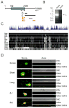
Figure 1. 3′UTR isoforms of importin β
(A) Schematic of the rat importin β1 transcript, with arrows indicating primers used for 3′RACE PCR. The blue box denotes the open reading frame. Lines under the schematic delineate regions subcloned for constructs containing long (L) or short (S) 3′UTR variants, or sequence regions used for deletion analysis (Δ1 and Δ2) of the long UTR. (B) Nested 3′RACE PCR on axonal and cell body cDNA reveals different 3′UTR variants for importin β1, with a 1.15 kb L variant preferentially sorted to axons. For sequences of the two main S and L isoforms please see Genbank accession number JX096837 and Supplementary Figure S1A. (C) Evaluation of importin β1 long 3′UTR conservation between species using the UCSC genome browser. The upper wiggle histogram summarizes conservation scores across 46 vertebrate genomes. Conserved sites with positive scores are shown in blue, while fast-evolving sites have negative scores. Pairwise alignments of 11 species to the human genome are displayed below the conservation histogram as a grey scale density plot, in which identity is shown in black. (D) Representative images of in situ hybridization for GFP riboprobe (red) and immunohistochemistry for neurofilament (green) on cell bodies (left) and distal axon shafts (right) of rat DRG cultures transfected with the indicated importin β1 3′UTR reporter constructs. The long 3′UTR and the Δ2 region both drive axonal localization of GFP mRNA while the short 3′UTR and the Δ1 region do not. Hybridization with sense GFP riboprobes shows no signal in cell body or axons for the long UTR. Scale bars: left = 30 μm, right = 20 μm.
Figure 2
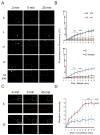
Figure 2. The long importin β1 3′UTR isoform confers axon localization
(A) Representative images from time-lapse sequences of fluorescence recovery after photobleaching (FRAP) experiments before (−2 min) and after photobleaching (0 and 20 min) of adult DRG neurons transfected with 3′ UTR constructs fused to myristylated GFP. The boxed regions indicate the area subjected to FRAP with recovery monitored over 20 min (for full time series please see Supplementary Figure S2A). Fluorescence recovery was observed only for the constructs containing the 3′ terminal segment of the long UTR isoform (L and Δ2) and this recovery was blocked upon incubation with the translation inhibitor anisomycin. Scale bar 25 μm. (B) Quantification of fluorescence recovery over multiple time-lapse sequences with the indicated constructs. Average recoveries are shown as % of pre-bleach levels ± SD. n ≥ 6 for each series, •• denotes p < 0.01, ••• denotes p < 0.001 (Two-way ANOVA). For quantification data for additional constructs please see Supplementary Figure S2B. (C) Representative images from time-lapse sequences of photo-conversion experiments before (−4 min) and after photo-conversion (0 and 40 min) of adult DRG neurons transfected with 3′ UTR constructs fused to myristylated Dendra2 (for full time series please see Supplementary Figure S3). Green represents unconverted or newly synthesized Dendra2; red shows photo-converted Dendra2. The boxed regions indicate the area subjected to a single round of laser-induced photo–conversion at a wavelength of 408 nm. The more proximal region was repetitively photo-converted to ensure that any green signal in the boxed region must arise from localized new synthesis of Dendra2. De novo synthesis of Dendra2 was observed for the Δ2 construct containing the 3′ end segment of the long UTR, but not for the short (S) UTR. Scale bar 25 μm. (D) Quantification of de novo Dendra2 synthesis from fluorescence intensity in the green channel over multiple time-lapse photoconversion sequences. Average ± SEM, n = 15. ** denotes p < 0.01 (Two-way ANOVA).
Figure 3
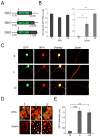
Figure 3. Validation of the importin β1 axon localization region in transgenic mice
(A) Schematic of DNA constructs used to generate transgenic mice. The neuronal specific Tα1 tubulin promoter was used to drive expression of a destabilized myristylated GFP fused to the 5′UTR of Importin β1, and different variants of Importin β1 3′UTR as indicated. (B) Quantitative RT-PCR for GFP mRNA from adult transgenic mouse tissue extracts. β actin served as an internal control. Left – GFP mRNA levels in DRG from the different transgenic lines, normalized to short 3′UTR line; Right – GFP mRNA levels in sciatic nerve from the different transgenic lines, normalized to DRG levels for each line. (C) Cultured adult sensory neurons from transgenic 3′UTR reporter mice (S, L, and Δ2 lines) after 48 hr in vitro. Robust GFP levels (green) are seen in neuronal cell bodies and in axons for the long and Δ2 UTR lines, but only in the cell body for the short 3′UTR line. Scale bar 20 μm, scale bar zoom panels 10 μm. (D) Merged images of GFP (green) and immunostaining for NF-H (red) on DRG and sciatic nerve sections from short, long and Δ2 transgenic lines seven days following sciatic nerve crush. For individual channel images please see Supplementary Figure S4. Axonal GFP signals are clearly observed in sciatic nerve sections from the long and Δ2 transgenic lines, but not from the short UTR line, despite equivalent expression in neuronal cell bodies in ganglia sections. Scale bar 10 μm. (E) Quantification of GFP intensity levels (au, arbitrary units) in sciatic nerve sections from short, long and 3′ terminal segment (Δ2) using CellProfiler image analysis software (average ± SEM) •• denotes p < 0.01, ••• denotes p < 0.001.
Figure 4
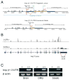
Figure 4. Conditional targeting of the axon-localizing region in importin β1 3′UTR
(A) Targeting strategy for importin β1 3′UTR. The loxP insertion sites are marked in red and the location of the 3′ and 5′ homology arms are in black. The PGK-neo selection cassette is inserted downstream of the region to be deleted (orange arrows) and flanked by FRT sites (green) that can be deleted using FLP recombinase. Three SV40 polyA signals are inserted immediately downstream of the floxed region (yellow boxes). For full sequences of the targeting construct and recombined locus please see Supplementary Procedures. (B) Deep sequencing analyses on RNA from knockout and wild type DRG confirms deletion of the targeted 3′UTR region in PGK-Cre/Importin β1 3′UTR loxP mice. (C) RT-PCR on RNA from knockout brain, DRG and sciatic nerve confirms complete deletion of the targeted 3′UTR region in PGK-Cre/Importin β1 3′UTR loxP mice.
Figure 5
Figure 5. The long 3′UTR is required for importin β1 localization to axons
(A) Quantification of relative importin β1 transcript levels in adult DRG and sciatic nerve extracts of wild type and PGK-Cre targeted mice. Note the significant decrease in message levels in the knockout nerve (residual message is likely from glial component of the tissue) coupled with increase in knockout DRG, consistent with accumulation of importin β1 transcript in ganglia due to the lack of an axon-localizing element. β actin served as an internal control, average ± SEM, n=4, ••• denotes p < 0.001 (unpaired two sample t-test). For additional analyses using Adv-Cre targeted mice, please see Supplementary Figure S5. (B) Representative images and quantification of Western blots for axoplasmic importin β1 in sciatic nerves of wild type and knockout animals before and 6 hours after injury, average ± SEM, n=3, * p < 0.05. Erk1/2 (ERK) was used as a loading control. (C) In situ hybridization for importin β1 (red) on NF-H positive (green) sensory neurons in culture. Scale bars - 20μm upper panels, 10 μm lower axon zoom panels. (D) Quantification of importin β1 mRNA in axons from neuronal cultures using CellProfiler image analysis software, average % intensity ± SEM, n=20, ** denotes p < 0.01. (E) In situ hybridization for importin β1 (red) on longitudinal sections from wild type or knockout sciatic nerve. Sensory axons are identified with NF-H (grey). Arrows mark axonal transcript, while arrowheads mark signal in adjacent Schwann cells. Scale bar 5 μm. (F) Quantification of importin β1 mRNA in axons from longitudinal sections, average % intensity ± SEM, n=3, ** p < 0.01. (G) Immunostaining for Importin β1 (red) and NF-H (green) on adult DRG neurons in culture. Scale bar 20 μm. (H) Quantification of Importin β1 immunoreactivity in axons from neuronal cultures using CellProfiler image analysis software, average % intensity ± SEM, n=20, •• denotes p < 0.01. (I) Cross-sections of sciatic nerve taken 6 hr after crush lesion and immunostained for importin β1 (red) and the neuronal marker NF-H (green). Scale bar 20 μm. (J) Quantification of axonal importin β1 immunoreactivity in the sciatic nerve cross sections using CellProfiler, average % intensity ± SEM, n=20, ••• denotes p < 0.001.
Figure 6
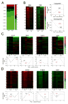
Figure 6. Lack of Axonal Importin β1 Attenuates the Cell Body Transcriptional Response to Nerve Injury
(A) Deep sequencing data-sets from RNA isolated from knockout or wild-type DRG were analyzed with DESeq. The heat maps shown were generated using FPKM (fragments per kilobase per million sequenced reads) values for 16,383 RefSeq genes expressed in the tissue. Two biological replicates were sequenced for each sample with essentially identical results, only 154 genes out of 27,563 differed between the genotypes. (B) Comparative L4/L5 DRG gene expression analyses between wild type and 3′UTR knockout mice after sciatic nerve lesion. RNA was extracted at three time points after injury (6, 12, and 18 hours) and analyzed using Illumina expression microarrays (n=3 for each sample). The heat maps show clusters of differentially expressed genes with log2 fold changes as indicated by the color key to the right. The traces on the right show the averaged trend of up- or down-regulation for the entire cluster for both genotypes. Numbers of genes in each cluster are indicated above the corresponding heat map. The two clusters shown in this panel are the largest (836 upregulated and 725 downregulated genes, respectively) that show highly significant (p ≤ 0.005, ANOVA) attenuation in their transcriptional regulation in knockout DRG as compared to wild type. (C) Additional and smaller clusters of differentially expressed genes that show highly significant (p ≤ 0.005) attenuation in their transcriptional regulation in knockout DRG as compared to wild type. Numbers of genes in each cluster are indicated above the corresponding heat map. (D) Clusters of differentially expressed genes that show significant (0.005 ≤ p ≤ 0.05) attenuation in their transcriptional regulation in knockout DRG as compared to wild type. Numbers of genes in each cluster are indicated above the corresponding heat map. Additional gene clusters regulated by injury that did not differ significantly between knockout and wild type are shown in Supplementary Figure S6. Gene-lists for all the clusters are provided in Supplementary Table S1.
Figure 7
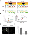
Figure 7. Lack of Axonal Importin β1 Delays Recovery from Sciatic Nerve Injury
(A) Representative images of CatWalk gait analysis (Noldus, version 9.0) with analyses of footprints and gait from wild type and PGK-Cre targeted mice before and six days after unilateral (right) sciatic nerve lesion. Six days after injury, the knockout is making less use of the right hind limb (ipsilateral to the injury) than the wild type (red arrow). (B) Quantification of recovery time course for print area in injured right hind limb. The print area of the ipsilateral hind paw is expressed in relation to the print area of the contralateral hind paw over subsequent days (in percent). Data are expressed as average ± SEM, n=12, * denotes p < 0.05, ** denotes p < 0.01 (Two-way ANOVA). For a complete time course up until Day 26 and more extensive statistical analyses please see Supplementary Figure S7A. (C) Quantification of recovery for the duty cycle parameter in injured right hind limb. The duty cycle of the ipsilateral hind paw is expressed in relation to the duty cycle of the contralateral hind paw over subsequent days (in percent). Data are expressed as average ± SEM, n=12, * denotes p < 0.05 (Two-way ANOVA). For a complete time course up until Day 26 and more extensive statistical analyses please see Supplementary Figure S7B. (D) YFP-expressing sensory axons after sciatic nerve lesion. Representative images of longitudinal sections 2 mm distal to the injury site, from YFP/wild type or YFP/knockout sciatic nerve, six days after sciatic lesion. Scale bar 100 μm. (E) Quantification of YFP levels in axonal fibers reveals significant differences between wild-type and importin β1 3′UTR null nerves six days after lesion. Average intensity ± SEM, n=6, ** denotes p < 0.01 (independent sample t-test).

Similar articles

Cited by

References

    1. Aakalu G, Smith WB, Nguyen N, Jiang C, Schuman EM. Dynamic visualization of local protein synthesis in hippocampal neurons. Neuron. 2001;30:489–502. - PubMed
    1. Abe N, Cavalli V. Nerve injury signaling. Curr Opin Neurobiol. 2008;18:276–283. - PMC - PubMed
    1. An JJ, Gharami K, Liao GY, Woo NH, Lau AG, Vanevski F, Torre ER, Jones KR, Feng Y, Lu B, et al. Distinct role of long 3′ UTR BDNF mRNA in spine morphology and synaptic plasticity in hippocampal neurons. Cell. 2008;134:175–187. - PMC - PubMed
    1. Anders S, Huber W. Differential expression analysis for sequence count data. Genome Biol. 2010;11:R106. - PMC - PubMed
    1. Andreassi C, Riccio A. To localize or not to localize: mRNA fate is in 3′UTR ends. Trends Cell Biol. 2009;19:465–474. - PubMed

Publication types

MeSH terms