

Stem-cell-based, tissue engineered tracheal replacement in a child: a 2-year follow-up study
- PMID: 22841419
- PMCID: PMC4487824
- DOI: 10.1016/S0140-6736(12)60737-5
Stem-cell-based, tissue engineered tracheal replacement in a child: a 2-year follow-up study
Abstract
Background: Stem-cell-based, tissue engineered transplants might offer new therapeutic options for patients, including children, with failing organs. The reported replacement of an adult airway using stem cells on a biological scaffold with good results at 6 months supports this view. We describe the case of a child who received a stem-cell-based tracheal replacement and report findings after 2 years of follow-up.
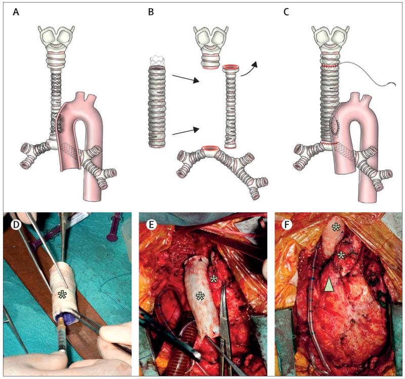
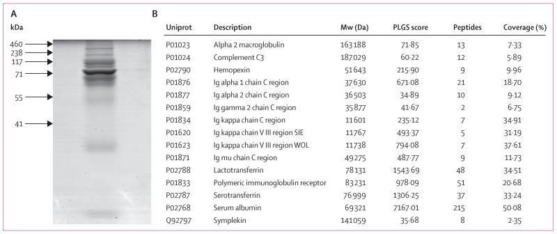
Methods: A 12-year-old boy was born with long-segment congenital tracheal stenosis and pulmonary sling. His airway had been maintained by metal stents, but, after failure, a cadaveric donor tracheal scaffold was decellularised. After a short course of granulocyte colony stimulating factor, bone marrow mesenchymal stem cells were retrieved preoperatively and seeded onto the scaffold, with patches of autologous epithelium. Topical human recombinant erythropoietin was applied to encourage angiogenesis, and transforming growth factor β to support chondrogenesis. Intravenous human recombinant erythropoietin was continued postoperatively. Outcomes were survival, morbidity, endoscopic appearance, cytology and proteomics of brushings, and peripheral blood counts.
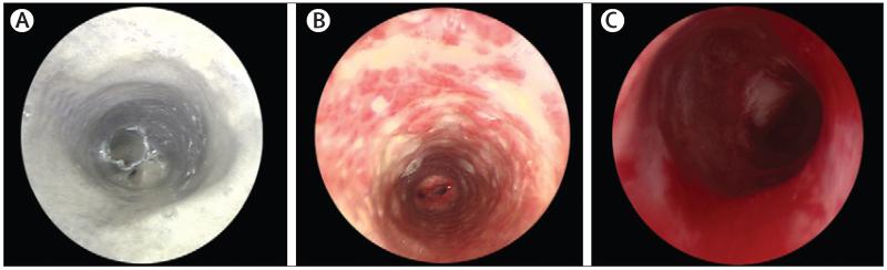
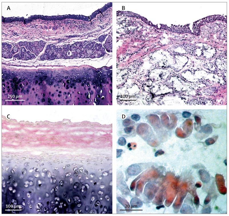
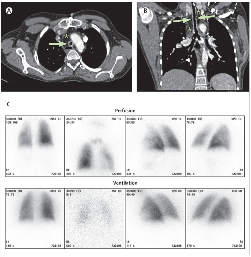
Findings: The graft revascularised within 1 week after surgery. A strong neutrophil response was noted locally for the first 8 weeks after surgery, which generated luminal DNA neutrophil extracellular traps. Cytological evidence of restoration of the epithelium was not evident until 1 year. The graft did not have biomechanical strength focally until 18 months, but the patient has not needed any medical intervention since then. 18 months after surgery, he had a normal chest CT scan and ventilation-perfusion scan and had grown 11 cm in height since the operation. At 2 years follow-up, he had a functional airway and had returned to school.
Interpretation: Follow-up of the first paediatric, stem-cell-based, tissue-engineered transplant shows potential for this technology but also highlights the need for further research.
Funding: Great Ormond Street Hospital NHS Trust, The Royal Free Hampstead NHS Trust, University College Hospital NHS Foundation Trust, and Region of Tuscany.
Copyright © 2012 Elsevier Ltd. All rights reserved.
Figures
Comment in
-
Engineering tissues for children: building grafts that grow.Lancet. 2012 Sep 15;380(9846):957-8. doi: 10.1016/S0140-6736(12)60951-9. Epub 2012 Jul 26. Lancet. 2012. PMID: 22841418 No abstract available.
-
Tracheal tissue engineering: building on a strong foundation.Expert Rev Med Devices. 2013 Jan;10(1):33-5. doi: 10.1586/erd.12.74. Expert Rev Med Devices. 2013. PMID: 23278221
-
Stem-cell-based, tissue-engineered tracheal replacement in a child.Lancet. 2013 Jan 12;381(9861):113. doi: 10.1016/S0140-6736(13)60043-4. Lancet. 2013. PMID: 23312748 No abstract available.
-
Stem-cell-based, tissue-engineered tracheal replacement in a child - Authors' reply.Lancet. 2013 Jan 12;381(9861):113. doi: 10.1016/S0140-6736(13)60044-6. Lancet. 2013. PMID: 23312749 No abstract available.
References
-
- Kocyildirim E, Kanani M, Roebuck D, et al. Long-segment tracheal stenosis: slide tracheoplasty and a multidisciplinary approach improve outcomes and reduce costs. J Thorac Cardiovasc Surg. 2004;128:876–82. - PubMed
-
- Jacobs JP, Quintessenza JA, Botero LM, et al. The role of airway stents in the management of pediatric tracheal, carinal and bronchial disease. Eur J Cardiothor Surg. 2000;18:505–12. - PubMed
-
- Lange P, Fishman JM, Elliott MJ, De Coppi P, Birchall MA. What can regenerative medicine offer for infants with laryngotracheal agenesis? Otolaryngol Head Neck Surg. 2011;145:544–50. - PubMed
-
- Atala A, Bauer SB, Soker S, Yoo JJ, Retik AB. Tissue-engineered autologous bladders for patients needing cystoplasty. Lancet. 2006;367:1241–46. - PubMed
-
- Macchiarini P, Jungebluth P, Go T, et al. Clinical transplantation of a tissue-engineered airway. Lancet. 2008;372:2023–30. - PubMed
Publication types
MeSH terms
Substances
Grants and funding
LinkOut - more resources
Full Text Sources
Other Literature Sources
