. 2012 Aug 14;23(2):433-40.
doi: 10.1016/j.devcel.2012.06.002. Epub 2012 Jul 26.

Disruption of microtubule integrity initiates mitosis during CNS repair

Affiliations

Disruption of microtubule integrity initiates mitosis during CNS repair

Torsten Bossing et al. Dev Cell. .

Abstract

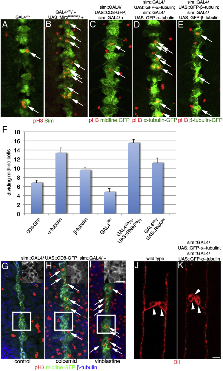
Mechanisms of CNS repair have vital medical implications. We show that traumatic injury to the ventral midline of the embryonic Drosophila CNS activates cell divisions to replace lost cells. A pilot screen analyzing transcriptomes of single cells during repair pointed to downregulation of the microtubule-stabilizing GTPase mitochondrial Rho (Miro) and upregulation of the Jun transcription factor Jun-related antigen (Jra). Ectopic Miro expression can prevent midline divisions after damage, whereas Miro depletion destabilizes cortical β-tubulin and increases divisions. Disruption of cortical microtubules, either by chemical depolymerization or by overexpression of monomeric tubulin, triggers ectopic mitosis in the midline and induces Jra expression. Conversely, loss of Jra renders midline cells unable to replace damaged siblings. Our data indicate that upon injury, the integrity of the microtubule cytoskeleton controls cell division in the CNS midline, triggering extra mitosis to replace lost cells. The conservation of the identified molecules suggests that similar mechanisms may operate in vertebrates.


Figures

Figure 1
Figure 1
Damage at the Ventral Midline in Drosophila Embryos Releases Midline Cells from G2 Arrest Genotypes listed at top of panels. Brackets indicate midline. Ventral views, anterior up. Scale, 6 μm. (A–D) Midline cells (green) in early stage 11 embryos do not divide (A). After mass removal of cells (arrowheads, B–D), adjacent midline cells enter into mitosis (arrows, B, D). Dividing cells are labeled with phosphoHistone H3 antibodies (red, pH3). Time in minutes after ablation. (C) and (D) show different time points of the same embryo. (E) One DiI-labeled midline precursor (red) in JupiterG00147 embryos (green) in which microtubules are outlined by GFP. All midline cells divide nearly simultaneously shortly after gastrulation. (F) After completion of the initial division, all midline cells (green, Single minded antibody) enter into S phase (red, BrdU). (G) At early stage 13 cyclinB (green) strongly accumulates in midline cells and a small subset of cells divide again (red). (H and H’) For 3.5 hr after the first division, midline cells (green) are devoid of string mRNA (red), which encodes for cdc25 phosphatase required for entry into mitosis. H’ shows only string mRNA. (I) Midline cells in early stage 11 do not express string mRNA (red). (J) string is transcribed in midline cells (arrows) after mechanical damage (arrowhead). (K–M) Time sequence of midline repair. Two siblings are generated by the initial division of one labeled midline precursor (red, DiI, K). Ablation of one sibling (arrowhead, K), results in division of the surviving sibling (L) and replacement of the lost sibling (bottom clone, M). A second labeled precursor in the same embryo was left undisturbed (top clone, M). Both precursors gave rise to a VUM clone consisting of one interneuron and one motorneurons. i, interneuronal projection; m, motorneuronal axon. (N) Ablations of midline siblings cause replacement of ablated sibling (green bar) or cell death (gray bar). Replacement always results in clonal types previously described in wild-type. Clonal types are: VUM, ventral unpaired median neurons; MP1, midline precursor 1 neurons; UMI, unpaired median interneurons; MNB, median neuroblast; MG, midline glia; undiff, two round cells with no differentiation. See also Figure S1 and Movies S1, S2, and S3.
Figure 2
Figure 2
Disruption of Microtubules Triggers Cell Divisions Genotypes listed at top of panel. Ventral views, anterior up. Bar, 6 μm. (A) In stage 13 embryos, a minority of midline cells (green) divide (arrows, red, pH3). (B) Depletion of Miro by expression of UAS::MiroRNAi in all tissues results in increased cell divisions in midline cells only. (C–E) Expression of GFP (green, C) does not increase mitosis (arrows, red, pH3) in stage 13 embryos but midline expression of α-tubulin (green, D) and, to a lesser degree, β-tubulin (green, E), triggers extra mitosis. (F) Number of dividing midline cells in all trunk segments of early stage 13 embryos. ANOVA, bars represent SEM. CD8::GFP, sim::GAL4/ UAS::CD8-GFP (n = 39)/ sim::GAL4/ +. α-tubulin, UAS::GFP-α−tubulin/ sim::GAL4; UAS::GFP-α-tubulin/ sim::GAL4 (n = 35). β-tubulin, UAS::GFP-β−tubulin/ sim::GAL4; UAS::GFP-β-tubulin/ sim::GAL4 (n = 36). GAL4V2h (n = 10). Gal4V2h/+; UAS::MiroRNATRIP02775/+ (n = 16). Gal4V2h/UAS::MiroRNAKK106683 (n = 23). Error bars: SEM. (G) In water-injected stage 10 embryos, midline cells (green) do not divide (red) and show a cortical accumulation of β-tubulin (blue). (H and I) Partial depolymerization of microtubules in stage 10 embryos injected with colcemid (n = 14) or vinblastine (n = 27) drives midline cells into division. Insets show higher magnification of boxed area and β-tubulin only. (I) Midline precursor 1 (MP1) in wild-type gives rise to two MP1-neurons (arrowheads). (J) Midline expression of α-tubulin results in extra MP1-neurons (arrowheads) generated by one precursor. See also Figures S2 and S3.
Figure 3
Figure 3
Cortical β-Tubulin Is Reduced by Mechanical Damage, Miro Depletion, and Expression of α-Tubulin Genotypes are listed on top of panels. Insets show higher magnifications of boxed area. A’–H’ shows tubulin only. Ventral views, anterior up. Bar, 6 μm. (A and A’) After mechanical damage, midline cells (green) at the wound (arrow) show a near loss of β-tubulin (red, arrows). (B and B’) Mechanical damage at the ventral midline (green) does not result in decreased α-tubulin staining (red) in cells at the damage site (arrows). Note, the antibody against β-tubulin recognizes mainly cortical tubulin whereas anti-α-tubulin outlines tubulin in cytoplasm and cortex. (C and C’) Midline cells show a robust staining against β-tubulin. (D and D’) Depletion of Miro by RNAi expression throughout the embryo results in a decrease in β-tubulin and an increased mitosis limited to the ventral midline. Depletion of Miro affects cortical tubulin but leaves the mitotic spindle and midbody (arrowhead) intact. (E–F’) In contrast to β-tubulin, the ubiquitous embryonic depletion of Miro (F, F’) leads to an increase of cytoplasmic α-tubulin (green) when compared to controls (E, E’). (G and G’) GFP expressing midline cells (green) show a robust β-tubulin stain. (H and H’) Expression of α-tubulin in midline cells reduces cortical β-tubulin but does not affect the spindle (arrowhead). See also Figure S4.
Figure 4
Figure 4
Jra, the Drosophila Jun Ortholog, Is Essential for Midline Repair Genotypes are listed on top of panels. Ventral views, anterior up. Bar 5 μm. (A and B) In heterozygous embryos (A), but not in Jra mutant embryos (B), removal of midline cells (green, arrowhead marks gap) triggers mitosis (arrows; pH3, red). (C) In heterozygous embryos, ablation of midline siblings leads to replacement of the ablated cell. (D) After ablation of one sibling in Jra mutants, the surviving sibling does not divide or differentiate. (E) Replacement of ablated sibling cells in heterozygous (JraIA109/Cyo, n = 21) and mutant (JraIA109/ JraIA109, n = 14) embryos. No repair, undamaged sibling does not divide; repair, undamaged sibling divides; dead, undamaged sibling dies. (F and G) At stage 13, in wild-type, jra mRNA (red) is not detectable in midline cells (green, F) but midline expression of α-tubulin activates jra transcription (red, G). Insets only show the red channel at the midline. See also Figure S4.

Comment in

References

    1. Arendt D., Nübler-Jung K. Comparison of early nerve cord development in insects and vertebrates. Development. 1999;126:2309–2325. - PubMed
    1. Bedelbaeva K., Snyder A., Gourevitch D., Clark L., Zhang X.M., Leferovich J., Cheverud J.M., Lieberman P., Heber-Katz E. Lack of p21 expression links cell cycle control and appendage regeneration in mice. Proc. Natl. Acad. Sci. USA. 2010;107:5845–5850. - PMC - PubMed
    1. Bergmann A., Tugentman M., Shilo B.Z., Steller H. Regulation of cell number by MAPK-dependent control of apoptosis: a mechanism for trophic survival signaling. Dev. Cell. 2002;2:159–170. - PubMed
    1. Bossing T., Technau G.M. The fate of the CNS midline progenitors in Drosophila as revealed by a new method for single cell labelling. Development. 1994;120:1895–1906. - PubMed
    1. Bossing T., Brand A.H. Determination of cell fate along the anteroposterior axis of the Drosophila ventral midline. Development. 2006;133:1001–1012. - PubMed

Publication types