. 2012 Oct;292(1-2):14-25.
doi: 10.1016/j.heares.2012.07.003. Epub 2012 Jul 25.

Immunogold TEM of otoconin 90 and otolin - relevance to mineralization of otoconia, and pathogenesis of benign positional vertigo

Affiliations

Immunogold TEM of otoconin 90 and otolin - relevance to mineralization of otoconia, and pathogenesis of benign positional vertigo

Leonardo R Andrade et al. Hear Res. 2012 Oct.

Abstract

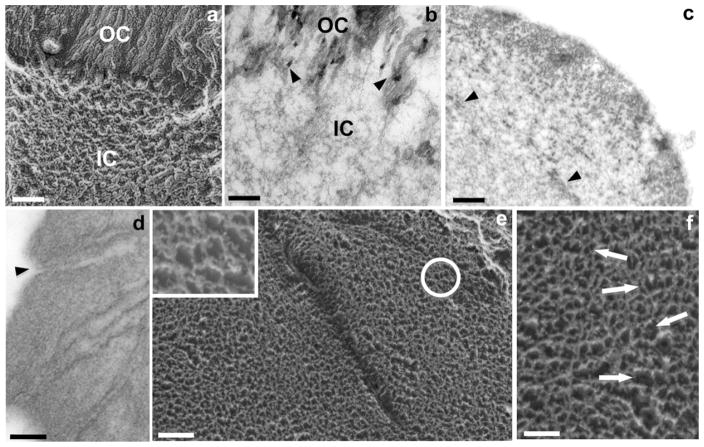
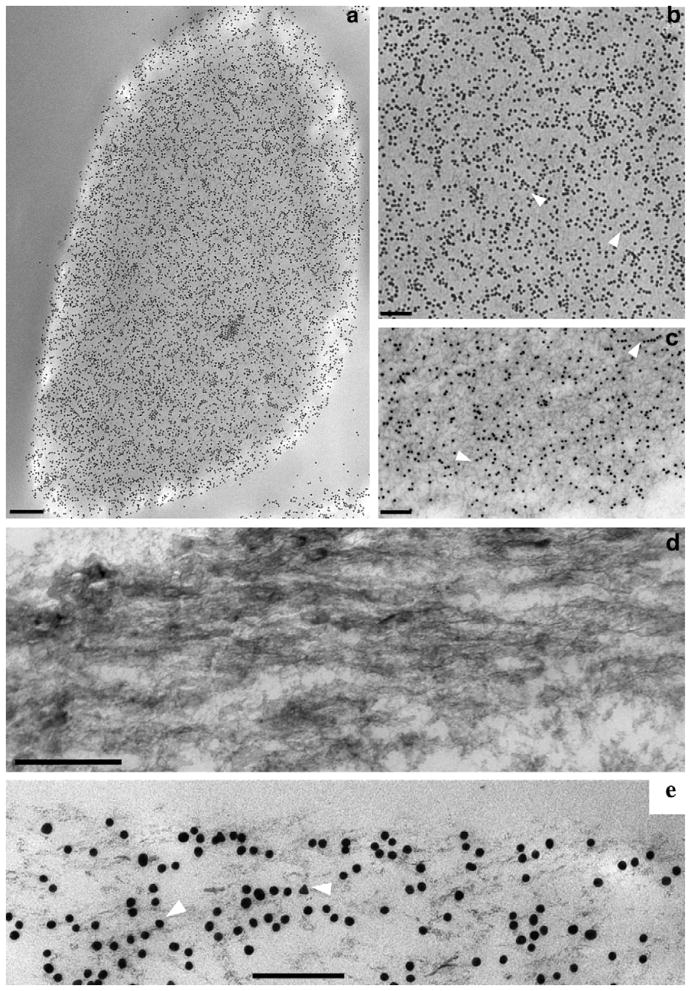
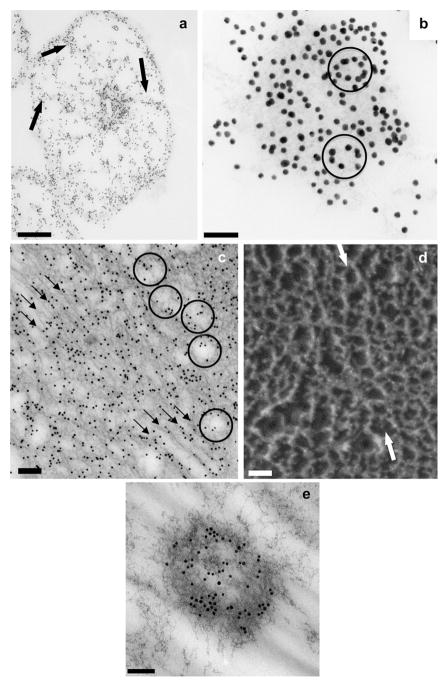
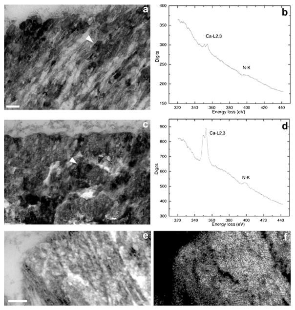
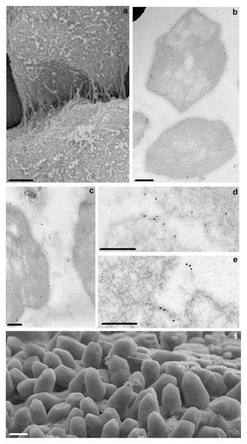
Implementation of the deep-etch technique enabled unprecedented definition of substructural elements of otoconia, including the fibrillar meshwork of the inner core with its globular attachments. Subsequently the effects of the principal soluble otoconial protein, otoconin 90, upon calcite crystal growth in vitro were determined, including an increased rate of nucleation, inhibition of growth kinetics and significant morphologic changes. The logical next step, ultrastructural localization of otoconin 90, by means of immunogold TEM in young mature mice, demonstrated a high density of gold particles in the inner core in spite of a relatively low level of mineralization. Here gold particles are typically arranged in oval patterns implying that otoconin 90 is attached to a scaffold consisting of the hexagonal fibrillar meshwork, characteristic of otolin. The level of mineralization is much higher in the outer cortex where mineralized fiber bundles are arranged parallel to the surface. Following decalcification, gold particles, as well as matrix fibrils, presumed to consist of a linear structural phenotype of otolin, are aligned in identical direction, suggesting that they serve as scaffold to guide mineralization mediated by otoconin 90. In the faceted tips, the level of mineralization is highest, even though the density of gold particles is relatively low, conceivably due to the displacement by the dense mineral phase. TEM shows that individual crystallites assemble into iso-oriented columns. Columns are arranged in parallel lamellae which convert into mineralized blocks for hierarchical assembly into the complex otoconial mosaic. Another set of experiments based on immunogold TEM in young mice demonstrates that the fibrils interconnecting otoconia consist of the short chain collagen otolin. By two years of age the superficial layer of mouse otoconia (corresponding to mid-life human) has become demineralized resulting in weakening or loss of anchoring of the fibrils interconnecting otoconia. Consequently, otoconia detached from each other may be released into the endolymphatic space by minor mechanical disturbances. In humans, benign positional vertigo (BPV) is believed to result from translocation of otoconia from the endolymphatic space into the semi-circular canals rendering their receptors susceptible to stimulation by gravity causing severe attacks of vertigo. The combinations of these observations in humans, together with the presented animal experiments, provide a tentative pathogenetic basis of the early stage of BPV.


Figures

Fig 1
Fig 1
Fine structural features of mammalian otoconia shown by different EM techniques. (a) SEM image of adult mouse otoconia showing typical barrel-shaped morphology and faceted ends; (b) freeze-etching of guinea pig otoconium showing pointed tip and rounded bodies with abundant filaments covering the surface (arrowhead); (c) TEM of a mouse otoconium showing both outer cortex (OC) and inner core (IC); (d) corresponding high magnification image showing fibrillar meshwork of inner core (IC) with several mineralized plaques (arrow), and outer cortex (OC) with parallel aligned mineralized fiber bundles (arrowhead); (e) freeze-etch image of cross-fractured guinea pig otoconium exposing parallel-aligned lamellae consisting of fused crystallites. On the right-hand side of the image, the rims of the cleavage planes exhibit sharp zigzagging patterns, representing the pointed tips of crystallites (arrowheads); (f) ultra-thin section of the densely mineralized cortex with parallel-arranged rows of iso-oriented columns of crystallites (arrowheads). The spaces between crystallites are widened due to sectioning procedure. Bars = 2 μm (a); 1 μm (b); 0.2 μm (c), 1 μm (d), 0.1 μm (e), 0.2 μm (f).
Fig 2
Fig 2
Fine structural features of the inner core of mammalian otoconia demonstrated by EM techniques. (a) Freeze-etching and (b) ultra-thin sectioning TEM; (b) images of the interface between inner core (IC) and outer cortex (OC) of otoconia. The freeze-etch image (a) demonstrates a rather abrupt border, but the ultra-thin section in (b) indicates that the fibrillar meshwork of the central core is continuous with the matrix of the outer cortex. The small dark plaques (arrowheads) in the cortex correspond to crystallites in the Bragg diffraction mode; (c) cross-section of otoconium showing electron-dense spots in inner core corresponding to mineralized matrix (arrowheads); (d) EDTA-treated otoconium showing several canals traversing outer cortex, connecting otoconial surface with inner core; (e) deep-etch image of freeze-fractured guinea pig otoconium showing the dense fibrillar web of inner core and innermost region of cortex. Inset: detail of area indicated by arrow showing typical hexagonal arrangement of fibrils; (f) high magnification of inner core of guinea pig otoconium showing several rows of co-aligned polygonal formations (arrows). Bars = 0.2 μm for (a) and (b); 0.3 μm (c); 0.5 μm (d); 0.2 μm (e) and 0.08 μm (inset); 0.1 μm (f).
Fig 3
Fig 3
Immunogold TEM of OC90 in outer cortex. (a) Low magnification view of longitudinal/tangential section of outer cortex, displaying high density of gold particles; (b) high magnification of Fig. 3a, demonstrating gold particles aligned in rows in straight or slightly curved patterns (arrowheads); (c) EDTA-treated cortical region of an otoconium, distinctly showing dense unstained fibrillar matrix as well as immunogold particles. In most regions gold particles are aligned in same direction as fibrils (arrowheads); (d) ultra-thin sagittal section of outer cortex of mouse otoconium; (e) corresponding section, immunogold labeled for OC90, indicating that the majority of the particles are aligned in the same direction of mineralized fibrils shown in (d) (white arrowheads). Bars = 0.15 μm (a); 0.2 μm (b); 0.2 μm (c); 0.1 μm (d); 0.1 μm (e).
Fig 4
Fig 4
Immunogold TEM of OC90 and deep-etch images of otoconia. (a) Cross-section through the equatorial plane of mouse otoconium showing strongly labeled inner core. Thin spoke-like projections radiate to periphery, merging with densely labeled subsurface layer (arrows); (b) high magnification image of central core showing several oval gold particle alignments (arrows) in addition to straight or slightly curved patterns; (c) longitudinal section of an EDTA-treated gold labeled inner core. Because of EDTA treatment, the unstained matrix is sharply contoured. Three rows of gold particles in the center of image delineate sets of diagonally oriented tubular formations corresponding to cavities in the fibrillar scaffold. Several diagonally oriented contiguous, co-aligned rows of oval/polygonal fibrillar formations are seen in periphery of image (arrow); (d) deep-etch image of freeze-fractured inner core, showing several rows of oval, co-aligned fibrillar formations (arrows) analogous to those seen in the thin sectioning TEM of Fig. 4c. [Note that two totally different techniques (thin sectioning TEM and freeze fracturing) show identical patterns.] (e) ectopic immunogold labeled structure resembling inner core on an otoconium, located in gelatinous membrane. Bars = 1 μm (a); 0.05 μm (b); 0.125 μm (c); 0.05 μm (d); 0.1 μm (e).
Fig 5
Fig 5
Analytical TEM demonstrates co-localization of mineral and organic phases. (a) TEM of the outer cortex of a mouse otoconium showing several small electron-dense mineral particles framed by fibrillar scaffolds (arrowhead); (b) electron energy loss spectrum of region indicated by arrowhead in (a), showing low peaks for both, calcium and nitrogen (markers for calcium carbonate and protein, respectively); (c) electron-micrograph of outer cortex showing a more advanced stage of mineralization than (a). The arrow points to the growing crystallites, which obscure fibrillar scaffold. The corresponding EELS shows a much higher calcium peak than (b), with the N peak unchanged (d); (e) elemental mapping of calcium of mouse otoconium in a zero-loss filtered image. Parallel columns of crystallites are separated by electron-lucent material (arrowhead); (f) calcium map of region depicted in (e). Bars = 0.1 μm for (a) and (c), 0.3 μm (e) and (f).
Fig 6
Fig 6
Interconnections of otoconia shown by SEM and immunogold TEM of otolin. (a) SEM image of adjacent otoconia showing dense fibrillar interconnections. The surface of PN3 day otoconia is covered with meandering fibrils whereas adjacent otoconia are interconnected by thin, tight fibrils; (b) and (c) images of ED18 mouse otoconia; (b) shows gold particles close to surface or within subsurface layer; (c) shows gold particles labeling interotoconial fibrils. (d) and (e) are images of young adult otoconia; (d) gold labeling of corresponding surfaces of otoconia; (e) pair of triplets of gold particles corresponding to interconnecting fibrils; (f) SEM image of otoconial layer of two-year-old mouse. Otoconial surface is denuded and otoconia are arranged in vertical orientation with tips projecting into endolymphatic lumen. Only lower tips of otoconia are loosely attached to the remnants of the fibrogelatinous matrix. Bars = 0.35 μm (a), 0.25 μm (b), 0.35 μm (c), 0.23 μm (d), 0.5 μm (e), 5.5 μm (f).

Similar articles

Cited by

References

    1. Agrawal Y, Carey JP, la Santina CC, Schubert MC, Minor LB. Disorders of balance and vestibular function in US adults: data from the National Health and Nutrition Examination Survey, 2001–2004. Archives of Internal Medicine. 2009;169:938–944. - PubMed
    1. Anniko M, Ylikoski J, Wroblewski R. Microprobe analysis of human otoconia. Acta Otolaryngology. 1984;97:283–289. - PubMed
    1. Baloh RW, Honrubia V, Jacobson K. Benign positional vertigo: clinical and oculographic features in 240 cases. Neurology. 1987;37:371–378. - PubMed
    1. Campos A, Canizares FJ, Sanchez-Quevedo MC, Romero PJ. Otoconial degeneration in the aged utricle and saccule. Advances in Otorhinolaryngology. 1990;45:143–153. - PubMed
    1. Colfen H. Biomineralization: a crystal-clear view. Nature Materials. 2010;9:960–961. - PubMed

Publication types