. 2012 Oct 1;370(1):110-24.
doi: 10.1016/j.ydbio.2012.07.017. Epub 2012 Jul 25.

Limb anterior-posterior polarity integrates activator and repressor functions of GLI2 as well as GLI3

Affiliations

Limb anterior-posterior polarity integrates activator and repressor functions of GLI2 as well as GLI3

Megan Bowers et al. Dev Biol. .

Abstract

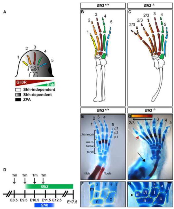
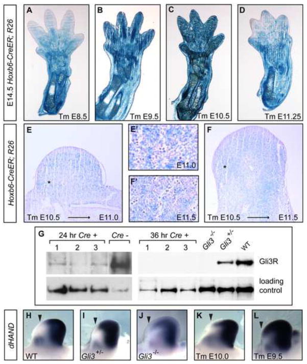
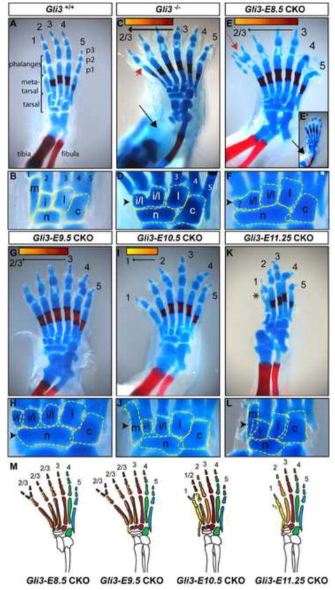
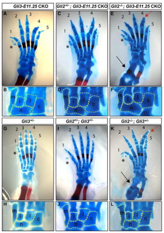
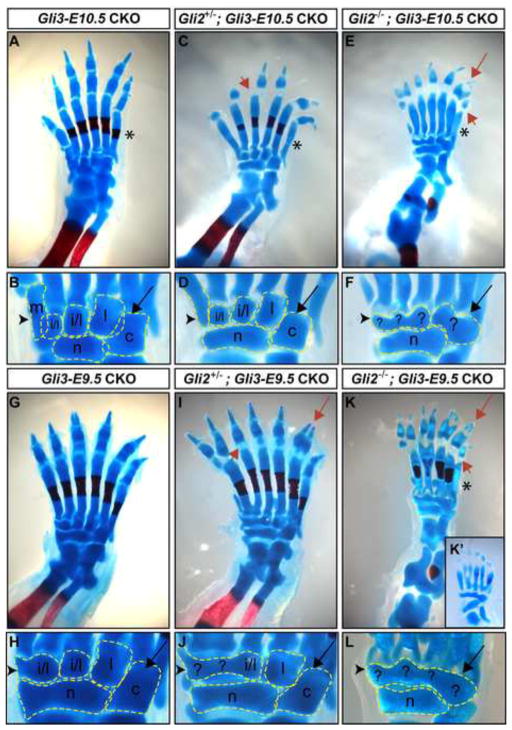
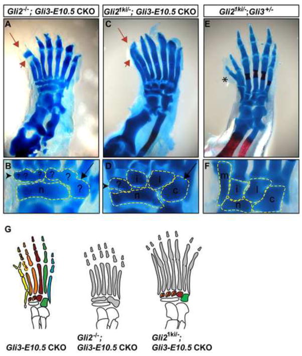
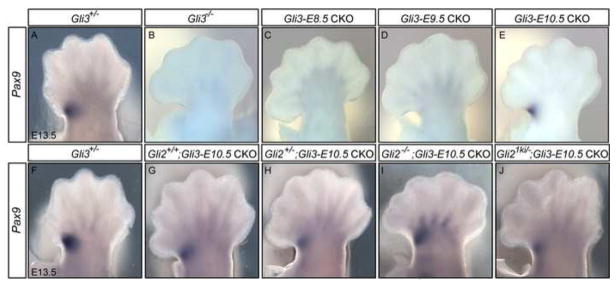
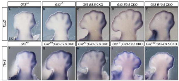
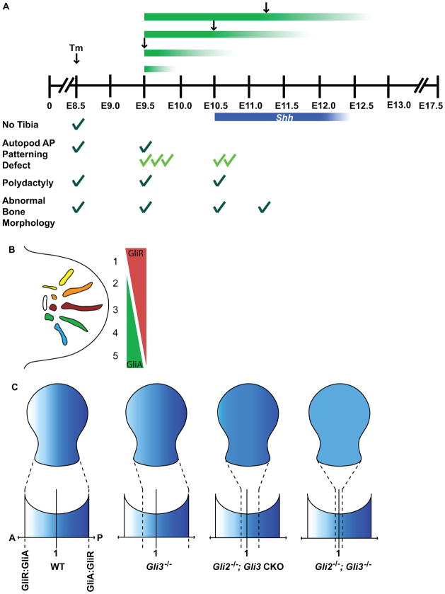
Anterior-posterior (AP) limb patterning is directed by sonic hedgehog (SHH) signaling from the posteriorly located zone of polarizing activity (ZPA). GLI3 and GLI2 are the transcriptional mediators generally utilized in SHH signaling, and each can function as an activator (A) and repressor (R). Although GLI3R has been suggested to be the primary effector of SHH signaling during limb AP patterning, a role for GLI3A or GLI2 has not been fully ruled out, nor has it been determined whether Gli3 plays distinct roles in limb development at different stages. By conditionally removing Gli3 in the limb at multiple different time points, we uncovered four Gli3-mediated functions in limb development that occur at distinct but partially over-lapping time windows: AP patterning of the proximal limb, AP patterning of the distal limb, regulation of digit number and bone differentiation. Furthermore, by removing Gli2 in Gli3 temporal conditional knock-outs, we uncovered an essential role for Gli2 in providing the remaining posterior limb patterning seen in Gli3 single mutants. To test whether GLIAs or GLIRs regulate different aspects of AP limb patterning and/or digit number, we utilized a knock-in allele in which GLI1, which functions solely as an activator, is expressed in place of the bifunctional GLI2 protein. Interestingly, we found that GLIAs contribute to AP patterning specifically in the posterior limb, whereas GLIRs predominantly regulate anterior patterning and digit number. Since GLI3 is a more effective repressor, our results explain why GLI3 is required only for anterior limb patterning and why GLI2 can compensate for GLI3A in posterior limb patterning. Taken together, our data suggest that establishment of a complete range of AP positional identities in the limb requires integration of the spatial distribution, timing, and dosage of GLI2 and GLI3 activators and repressors.


Figures

Fig. 1
Fig. 1
Gli3−/− hindlimbs develop polydactyly and severe AP patterning defects only in the anterior zeugopod and autopod. (A) Schematic of AP patterning mediated by Shh signaling. Grey shading represents a gradient of Shh signaling. (B,C) Schematic representations of wild type and Gli3−/− hindlimbs with each digit identity represented by a different color. Dotted lines denote bones that are variably lost. (D) Experimental design for CKO study showing when Tm was administered relative to Gli3 expression (green) and Shh expression (blue) in the hindlimb. (E,F) Wild type E17.5 hindlimb. (G,H) Gli3−/− hindlimbs develop digit polydactyly, with variable phalangeal bone duplications, variable abnormal p1 differentiation (red arrow-head), tibial agenesis (black arrow) and posteriorization of structures anterior to digit 3, including loss of digit 1 metatarsal morphology and ossification center, loss of digit 1 tarsal bone (black arrowhead) and anterior elongation of the navicular bone. Colored bar at the top of panel (G) indicates that digit and tarsal bones anterior to digit 3 adopt progressively more anterior pattern, but exclude digit 1 pattern. For all panels anterior is left, posterior right. m: medial cuneiform, i: intermediate cuneiform, l: lateral cuneiform, c: cuboid, n: navicular.
Fig. 2
Fig. 2
Hoxb6-CreER mediated recombination efficiency in the hindlimb mesenchyme. (A–F) Xgal staining in cross-sections of Hoxb6-CreER: R26R hindlimbs in embryos exposed to Tm at indicated stages and harvested at E14.5 (A–D), 36 hours post-gavage (E) and 48 hours post-gavage (F). Insets E′ and F′ are taken from regions of E and F indicated by *, respectively. (G) Western blots show GLI3R and control protein from hindlimb bud lysates of different single Gli3-E9.5 CKO mutants (Cre+, numbered) and sibling (Cre−) control embryos with genotypes as indicated, collected at 24 hours or at 36 hours after Tm treatment. Vinculin detection was used as a loading control. In the 24 hour blot, the Gli3fl/−; Cre negative sibling control sample lane was spliced to remove irrelevant lanes. The 24 hour and 36 hour examples shown are representative of results of 3 independent experiments (total of 7 mutant embryos analyzed at 24 hours post Tm treatment and 5 mutants analyzed at 36 hours post Tm treatment). (H–L) dHand in situs of E11.5 limb buds with indicated genotypes. The dHand domain is expanded anteriorly in Gli3−/− (n=10/10) hindlimbs compared to Gli3+/− (n=8/8). Anterior expansion is partial in Gli3 CKO limbs 36 hours post-gavage (6/8) and complete within 48 hours (7/8).
Fig. 3
Fig. 3
Hoxb6-CreER mediated Gli3 conditional knockout in the hindlimb reveals temporal-specific roles for Gli3 during limb patterning. The autopod shown in inset (E′) is an example of a Gli3-E8.5 CKO hindlimb with tibial bone loss (black arrow) similar to Gli3−/−. Black arrowhead draws attention to the anterior tarsal bone phenotype, which suggests digit 1 identity in J and L (compare to B), and correlates with the absence of digit 1 identity in D, F, and H. Note the retention of wild-type morphology in the cuboid and lateral cuneiform bones in all Gli3 mutants. (M) Schematic representation of E17.5 Gli3 CKO hindlimbs in which each digit identity is represented by a different color. The appearance of dislocation of the distal fibula and tibia in (E) and (K) is a consequence of tissue processing rather than a mutant phenotype. The appearance of digit 4 and 5 truncations in (E) and (K) is due to the angle of the photo, and is not a mutant phenotype. Red arrow: example of phalangeal duplication. Asterix: preaxial partial digit 1 duplication typical of Gli3+/− hindlimbs.See Fig. 1 for additional symbols.
Fig. 4
Fig. 4
Gli2 is not required for AP autopod patterning when Gli3 is removed by ~E13. (A–F) Both digit number and AP autopod patterning are normal in all Gli3-E11.25 CKO limbs, with and without Gli2. (G–L) Controls demonstrating pre-axial partial digit 1 duplication (asterix), digit 4 p3 duplication (red arrow) and shortened zeugopods bones (black arrow) typical of Gli3+/− (G,H), Gli2+/−; Gli3+/− (I,J) and Gli2−/−; Gli3+/− (K,L) hindlimbs. See Figs. 1 and 2 for additional symbols.
Fig. 5
Fig. 5
GLI2 provides AP patterning in autopods lacking Gli3 after E11.0. (A,B) Gli3-E10.5 CKO autopods develop polydactyly but retain a complete complement of normal digit identity. (C, D) Posteriorization of the anterior autopod of Gli2+/−; Gli3-E10.5 CKO mutants is suggested by lengthening of the first metatarsal bone and failure of the medial cuneiform bone to completely form. (E,F) Complete posteriorization of the anterior autopod and loss of posterior autopod patterning occurs in Gli2−/−; Gli3-E10.5 CKOs. (G, H) Gli3-E9.5 CKOs and (I, J) Gli2+/−; Gli3-E9.5 CKOs have similar anterior autopod patterning phenotypes. (K, L) Loss of anterior and posterior autopod patterning is evident in Gli2−/−; Gli3-E9.5 CKO tarsal bones. (K′) A Gli2−/−; Gli3-E9.5 CKO exhibiting nearly complete loss of AP polarity. Black arrowhead draws attention to the anterior tarsal bone of each mutant. Black arrow points to the two posterior tarsal bones. Red arrowhead: examples of abnormal p1 differentiation. Red arrow: examples of phalangeal duplications. Asterix: lack of complete metatarsal ossification center staining. For all panels anterior is left, posterior right. m: medial cuneiform, i: intermediate cuneiform, l: lateral cuneiform, c: cuboid, n: navicular.
Fig. 6
Fig. 6
GLIR patterns the anterior autopod and the posterior autopod is patterned by both GLIR and GLIA. (A,B) Gli2−/−; Gli3-E10.5 CKO autopods develop severely posteriorized anterior tarsal bones (black arrowhead) and anteriorized posterior tarsal bones (black arrow). (C,D) Gli21ki/−; Gli3-E10.5 CKO autopods develop severely posteriorized anterior tarsal bones, nearly normal posterior tarsal bone morphology. (E,F) Gli21ki/−; Gli3+/− are similar to Gli3+/− but have a more complete digit 1 duplication (asterix). Red arrowhead refers to loss of p1 in A and a gain-of-function phenotype associated with the Gli21ki allele, which causes the metatarsal bones to appear fused to the phalanges in C. Red arrow indicates examples of phalangeal duplications. (G) Schematic of autopod tarsal bone patterning in Gli CKOs.
Fig. 7
Fig. 7
Pax9 expression is restored in Gli3-E10.5 CKO limbs and correlates with the lack of AP polarity in Gli2; Gli3 CKO double mutant limbs. Dorsal view of Pax9 RNA in situs in E13.5 hindlimbs of the genotypes indicated. (A) Pax9 is expressed in the anterior proximal autopod in Gli3+/− limbs. Anterior Pax9 is lacking in Gli3−/− (B), Gli3-E8.5 CKO (C) and Gli3-E9.5 CKOs (D). Pax9 is expressed in Gli3-E10.5 CKOs, although reduced in some mutants (E, G). Pax9 appears reduced in some Gli2+/−; Gli3-E10.5 CKO hindlimbs (H) and in Gli21ki/−; Gli3-E10.5 CKOs (J), but is posteriorly expanded in some Gli2−/−; Gli3-E10.5 CKOs (I). Anterior is to the left in all images.
Fig. 8
Fig. 8
Tbx2 expression is expanded anteriorly in Gli3 conditional mutant limbs. (A) In E12.5 Gli3+/− hindlimbs, Tbx2 is detected in the anterior mesenchyme posterior to digit 3 and in the mesenchyme proximal to the AER posterior to digit 1. (B) Tbx2 expression is expanded anteriorly in Gli3−/− hindlimbs, posterior expression and distal mesenchymal expression is maintained. Similar anterior expansion of Tbx2 is seen in Gli3-E8.5 CKO (C), Gli3-E9.5 CKO (D) and Gli3-E10.5 CKO hindlimbs (E). In Gli2+/−; Gli3-E9.5 CKO (H) and Gli21ki/−; Gli3-E9.5 (J) CKO hindlimbs Tbx2 expression is expanded similar to in Gli3-E9.5 CKOs (G), but more extensively in Gli2−/−; Gli3-E9.5 CKOs (I). Anterior is to the left in all images.
Fig. 9
Fig. 9
Summary and model for GLI function in limb AP patterning. (A) Temporal conditional inactivation of Gli3 reveals the sequence of Gli3-mediated events during limb patterning. Removal of Gli2 in Gli3 CKOs reveals the requirement for Gli2 during AP patterning. Removal of Gli3 at sequentially earlier stages in Gli2 mutants emphasizes the combined role of duration and dosage of total GLI function to AP patterning. Black arrows demarcate the embryonic stage when Tm was administered. Green and blue bars indicate the expected duration of Gli3 and Shh expression, respectively. Dark green checks indicate phenotypes observed in Gli3 mutants, light green checks indicate phenotypes observed in Gli2; Gli3 CKOs. The number of checks represents the severity of each phenotype listed. (B) AP patterning of the anterior autopod is dependent on GliRs (red), corresponding to the spatial domain containing predominantly GliRs. AP patterning of the posterior autopod requires both GliA (green), and GliR function, which corresponds to the spatial domain of strong GliA function (Ahn and Joyner, 2004). (C) Polarity of the limb is altered in different Gli-mutant backgrounds. The range of polarity is decreased in Gli3−/− limbs, excluding the anterior-most positional identities. Removal of Gli2 in Gli3 CKO mutants further restricts the range of polarity so that distinct posterior positional identities are lost, and the morphology of tarsal bones converge upon a state that could represent the consequence of GliR:GliA=1. This predicts that a hypothetical Gli2−/−; Gli3−/− limb would completely lack AP polarity. Blue shading represents the range of positional identities present in a wild type limb. Dotted lines indicate the range of positional identities available in each genetic background.

References

    1. Ahn S, Joyner AL. Dynamic changes in the response of cells to positive hedgehog signaling during mouse limb patterning. Cell. 2004;118:505–16. - PubMed
    1. Bai CB, Joyner AL. Gli1 can rescue the in vivo function of Gli2. Development. 2001;128:5161–72. - PubMed
    1. Bai CB, Stephen D, Joyner AL. All mouse ventral spinal cord patterning by hedgehog is Gli dependent and involves an activator function of Gli3. Dev Cell. 2004;6:103–15. - PubMed
    1. Barna M, Pandolfi PP, Niswander L. Gli3 and Plzf cooperate in proximal limb patterning at early stages of limb development. Nature. 2005;436:277–81. - PubMed
    1. Bastida MF, Ros MA. How do we get a perfect complement of digits? Curr Opin Genet Dev. 2008;18:374–80. - PubMed

Publication types

MeSH terms