. 2012 Nov 15;318(19):2427-37.
doi: 10.1016/j.yexcr.2012.07.009. Epub 2012 Jul 27.

Molecular signature and in vivo behavior of bone marrow endosteal and subendosteal stromal cell populations and their relevance to hematopoiesis

Affiliations

Molecular signature and in vivo behavior of bone marrow endosteal and subendosteal stromal cell populations and their relevance to hematopoiesis

Alex Balduino et al. Exp Cell Res. .

Abstract

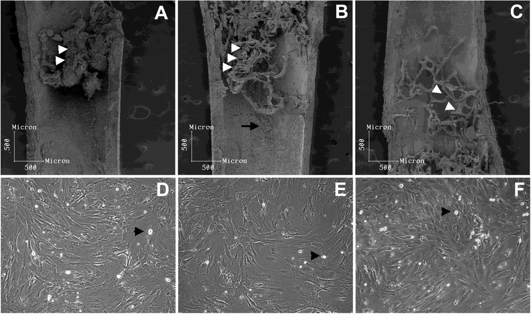
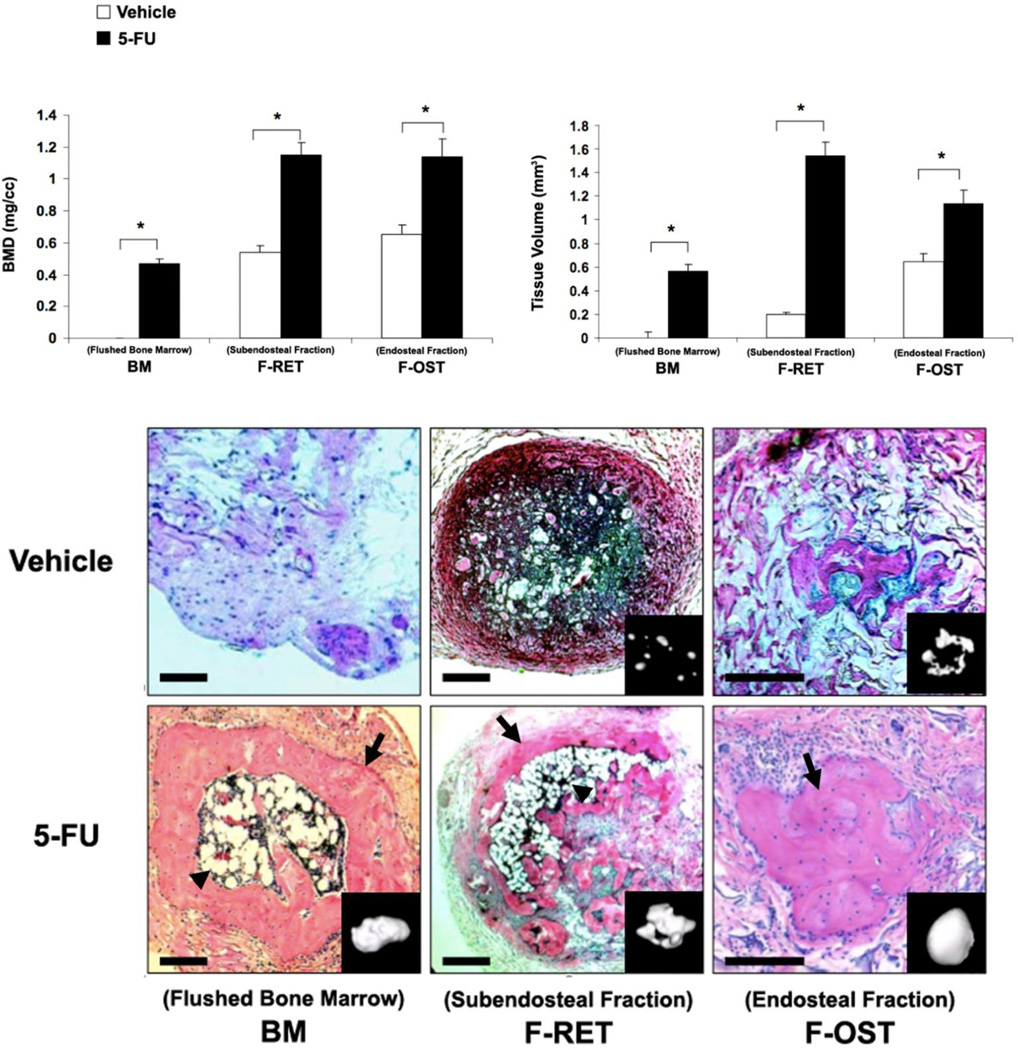
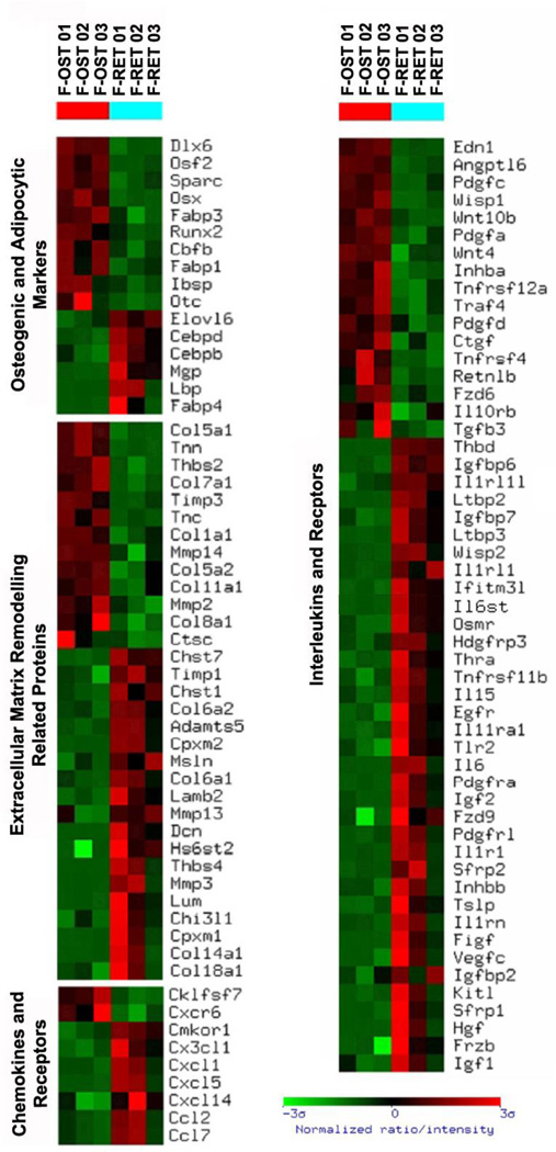
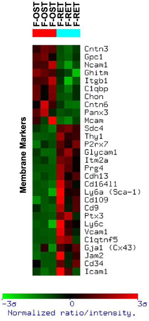
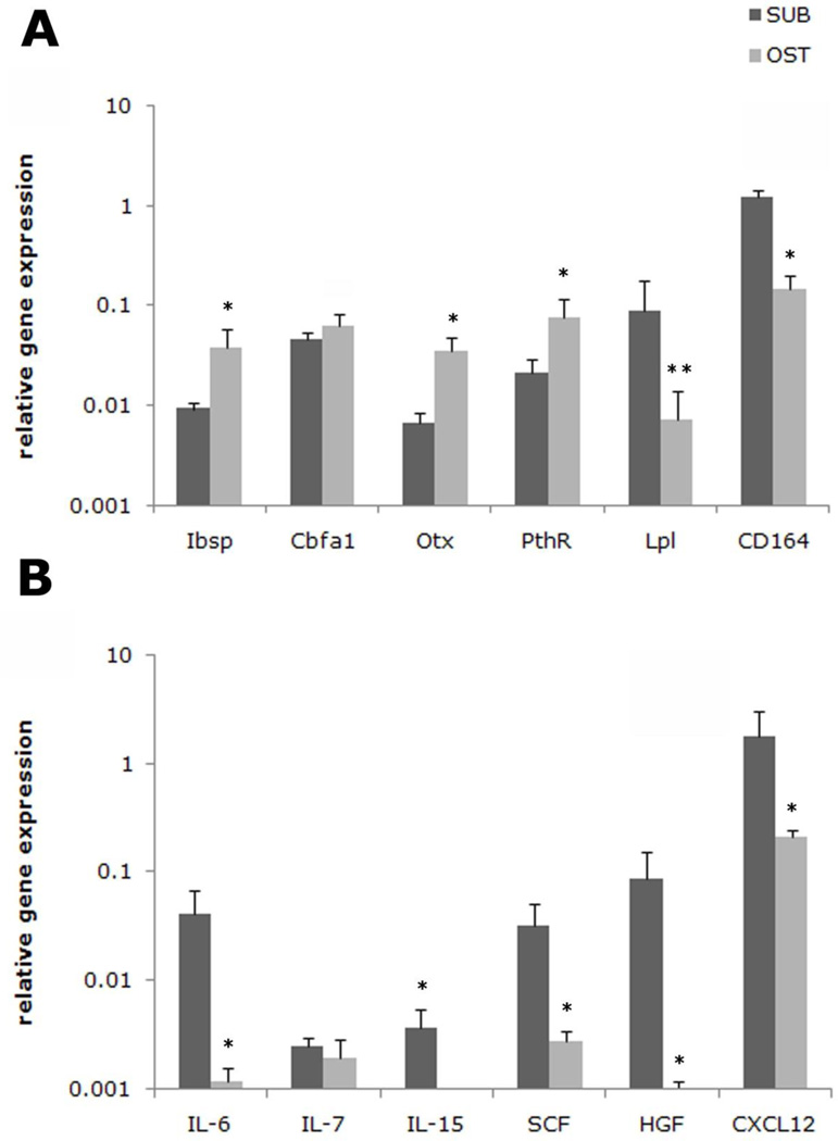
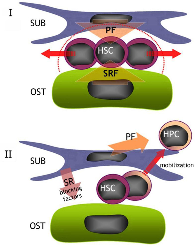
In the bone marrow cavity, hematopoietic stem cells (HSC) have been shown to reside in the endosteal and subendosteal perivascular niches, which play specific roles on HSC maintenance. Although cells with long-term ability to reconstitute full hematopoietic system can be isolated from both niches, several data support a heterogenous distribution regarding the cycling behavior of HSC. Whether this distinct behavior depends upon the role played by the stromal populations which distinctly create these two niches is a question that remains open. In the present report, we used our previously described in vivo assay to demonstrate that endosteal and subendosteal stromal populations are very distinct regarding skeletal lineage differentiation potential. This was further supported by a microarray-based analysis, which also demonstrated that these two stromal populations play distinct, albeit complementary, roles in HSC niche. Both stromal populations were preferentially isolated from the trabecular region and behave distinctly in vitro, as previously reported. Even though these two niches are organized in a very close range, in vivo assays and molecular analyses allowed us to identify endosteal stroma (F-OST) cells as fully committed osteoblasts and subendosteal stroma (F-RET) cells as uncommitted mesenchymal cells mainly represented by perivascular reticular cells expressing high levels of chemokine ligand, CXCL12. Interestingly, a number of cytokines and growth factors including interleukin-6 (IL-6), IL-7, IL-15, Hepatocyte growth factor (HGF) and stem cell factor (SCF) matrix metalloproteases (MMPs) were also found to be differentially expressed by F-OST and F-RET cells. Further microarray analyses indicated important mechanisms used by the two stromal compartments in order to create and coordinate the "quiescent" and "proliferative" niches in which hematopoietic stem cells and progenitors reside.


Conflict of interest statement

Authorship and Conflict of Interest Statements

Alex Balduino - designed research; performed research; analyzed data; wrote paper

Valeria Mello Coelho - designed research; analyzed data; wrote paper

Zhou Wang – performed research; analyzed data; wrote paper

Russell S. Taichman – designed research; analyzed data; wrote paper

Paul H. Krebsbach – analyzed data; wrote paper

Ashani T. Weeraratna - designed research; performed research; analyzed data

Kevin G. Becker - designed research; performed research; analyzed data

Wallace de Mello - performed research; analyzed

Dennis D. Taub - designed research; analyzed data; wrote paper

Radovan Borojevic - designed research; analyzed data; wrote paper

Conflict of interest statement

The authors declare no competing financial or potential conflict of interests.

Figures

Figure 1
Figure 1. Analysis of the inner surface of femurs and morphology of the stromal populations isolated
After bone marrow was flushed, subendosteal and endosteal stromal cells which remained attached were mainly distributed in the trabecular region (A, white arrow heads) at the metaphysis and epiphysis. After the first round of collagenase digestion procedure, subendostal stromal cells were removed (B) and osteoblasts remained attached (B, white arrow heads and arrow), being retrieved only after the second collagenase digestion (C, white arrow heads pointing to osteoblast-free trabecular bone). When in culture, flushed bone marrow (D) and subendosteal stromal cells (E) presented similar myofibroblastic morphology. Conversely, endosteal osteoblats (F) presented a cuboidal morphology, as expected. A few macrophage-like cells were observed in all three primary cultures (arrow heads). Primary cells were cultured to confluence in DMEM 10% FBS.
Figure 2
Figure 2. Histological and morphometric analyses of stromal cells fractions implants
As described in the Materials and Methods, 2×10⁶ of flushed bone marrow cells (BM), subendosteal stroma (F-RET), and endosteal osteoblasts (F-OST) isolated from normal and 5-FU treated mice were mixed up in gelatin sponge and transplanted into SCID mice subcutaneously. After 5 weeks, the implants were harvested for micro-CT scanning (inserts) and sectioned for histological staining using hematoxylin and eosin of the BM, F-FRET and F-OST tissues (lower panels). Original Magnification of 400×. Bone mass density (BMD) and tissue volume were measured (upper panels).
Figure 3
Figure 3. Gene expression profile based on gene function and physiology of subendosteal stromal cells (F-RET) compared to endosteal osteoblasts (F-OST)
Functional clustering was achieved and gene groups of particular interest are shown. The profile was created using the Ingenuity program. The brighter the red, the higher the gene expression level observed. The brighter the green, the lower the gene expression level observed. Only gene expression differences statistically significant are shown.
Figure 4
Figure 4. Gene expression profile of membrane-associated molecules of subendosteal stromal cells (F-RET) compared to endosteal osteoblasts (F-OST)
Functional clustering was achieved and gene groups of particular interest are shown. The profile was created using the Ingenuity program. The brighter the red, the higher the gene expression level observed. The brighter the green, the lower the gene expression level observed. Only gene expression differences statistically significant are shown.
Figure 5
Figure 5. Differences in relevant HSC niche gene expression by subendosteal stromal cells (F-RET) compared to endosteal osteoblasts (F-OST)
Real time RT-PCR analysis was performed on the subendosteal cells (SUB) and osteoblasts (OST) for the following genes identified using the microarray profiles: (A) Bone sialoprotein (Ibsp), Cbfa-1, osterix (Otx), Pth receptor (PthR), lipoprotein lipase (LPL) and CD164 and (B) interleukin 6 (IL-6), interleukin 7 (IL-7), interleukin 15 (IL-15), stem cell factor (SCF), hepatocyte growth factor (HGF), and stromal derived factor-1α (CXCL12) are presented. The results were normalized to GAPDH, which was also used to determine relative gene expression (dCT). Primers used are listed on Table 1.
Figure 6
Figure 6. Potential differences and roles for endosteal osteoblasts (F-OST) and subendosteal stromal cells (F-RET) in maintaining the HSC niche in the bone marrow
Our hypothesis says that, in the HSC niche, endosteal osteoblasts (F-OST) may supply the specific signals for self-renewal and quiescence (SRF). However, subendosteal stromal cells (F-RET) secrete growth factors and chemokines (PF) that, in a balance with stemness inducing factors secreted by osteoblasts, will promote HSC pool expansion (I). Conversely, subendosteal stromal cells (F-RET) secrete factors (SR blocking factors) which neutralize self-renewal factors presented by endosteal osteoblasts (F-OST), promoting HSC exit from the niche, proliferation and migration towards the central space.

Similar articles

Cited by

References

    1. Arai F, Hirao A, Ohmura M, Sato H, Matsuoka S, Takubo K, Ito K, Koh GY, Suda T. Tie2/angiopoietin-1 signaling regulates hematopoietic stem cell quiescence in the bone marrow niche. Cell. 2004;118:149–161. - PubMed
    1. Balduino A, Hurtado SP, Frazão P, Takiya CM, Alves LM, Nasciutti LE, El-Cheikh MC, Borojevic R. Bone marrow subendosteal microenvironment harbours functionally distinct haemosupportive stromal cell populations. Cell Tissue Res. 2005;319:255–266. - PubMed
    1. Bourke VA, Watchman CJ, Reith JD, Jorgensen ML, Dieudonnè A, Bolch WE. Spatial gradients of blood vessels and hematopoietic stem and progenitor cells within the marrow cavities of the human skeleton. Blood. 2009;114:4077–4080. - PMC - PubMed
    1. Brew K, Dinakarpandian D, Nagase H. Tissue inhibitors of metalloproteinases: evolution, structure and function. Biochim Biophys Acta. 2000;1477:267–283. - PubMed
    1. Butler JM, Nolan DJ, Vertes EL, Varnum-Finney B, Kobayashi H, Hooper AT, Seandel M, Shido K, White IA, Kobayashi M, Witte L, May C, Shawber C, Kimura Y, Kitajewski J, Rosenwaks Z, Bernstein ID, Rafii S. Endothelial cells are essential for the self-renewal and repopulation of Notch-dependent hematopoietic stem cells. Cell Stem Cell. 2010;6:251–264. - PMC - PubMed

Publication types

MeSH terms