

The effects of morphine on basal neuronal activities in the lateral and medial pain pathways
- PMID: 22841696
- PMCID: PMC3465732
- DOI: 10.1016/j.neulet.2012.07.032
The effects of morphine on basal neuronal activities in the lateral and medial pain pathways
Abstract
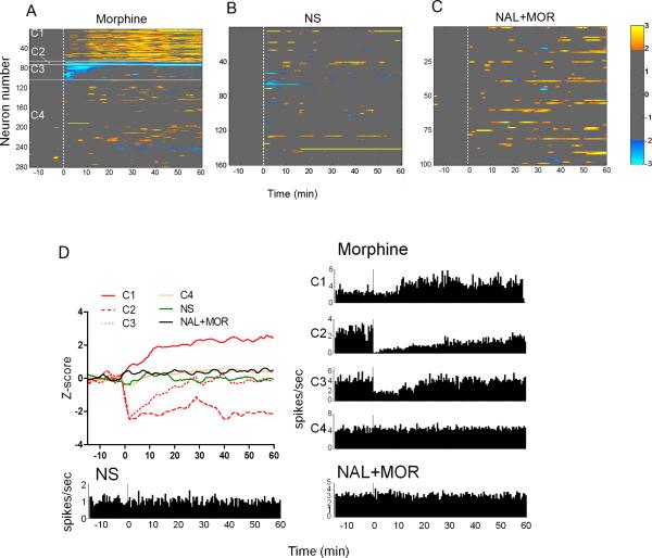
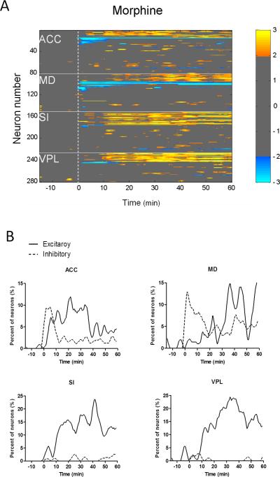
Numerous studies indicate that morphine suppresses pain-evoked activities in both spinal and supraspinal regions. However, little is known about the effect of morphine on the basal brain activity in the absence of pain. The present study was designed to assess the effects of single-dose morphine on the spontaneous discharge of many simultaneously recorded single units, as well as their functional connections, in the lateral pain pathway, including the primary somatosensory cortex (SI) and ventral posterolateral thalamus (VPL), and medial pain pathway, including the anterior cingulate cortex (ACC) and medial dorsal thalamus (MD), in awake rats. Morphine (5mg/kg) was administered intraperitoneally before the recording. Naloxone plus morphine and normal saline injections were performed respectively as controls. The results showed that morphine administration produced significant changes in the spontaneous neuronal activity in more than one third of the total recorded neurons, with primary activation in the lateral pathway while both inhibition and activation in the medial pathway. Naloxone pretreatment completely blocked the effects induced by morphine. In addition, the correlated activities between and within both pain pathways was exclusively suppressed after morphine injection. These results suggest that morphine may play different roles in modulating neural activity in normal vs. pain states. Taken together, this is the first study investigating the morphine modulation of spontaneous neuronal activity within parallel pain pathways. It can be helpful for revealing neuronal population coding for the morphine action in the absence of pain, and shed light on the supraspinal mechanisms for preemptive analgesia.
Copyright © 2012 Elsevier Ireland Ltd. All rights reserved.
Figures
Similar articles
-
Effects of pentobarbital anesthesia on nociceptive processing in the medial and lateral pain pathways in rats.Neurosci Bull. 2010 Jun;26(3):188-96. doi: 10.1007/s12264-010-0150-x. Neurosci Bull. 2010. PMID: 20502496 Free PMC article.
-
Morphine modulation of pain processing in medial and lateral pain pathways.Mol Pain. 2009 Oct 13;5:60. doi: 10.1186/1744-8069-5-60. Mol Pain. 2009. PMID: 19822022 Free PMC article.
-
Spontaneous Cingulate High-Current Spikes Signal Normal and Pathological Pain States.J Neurosci. 2019 Jun 26;39(26):5128-5142. doi: 10.1523/JNEUROSCI.2590-18.2019. Epub 2019 Apr 25. J Neurosci. 2019. PMID: 31023834 Free PMC article.
-
Functional imaging of brain responses to pain. A review and meta-analysis (2000).Neurophysiol Clin. 2000 Oct;30(5):263-88. doi: 10.1016/s0987-7053(00)00227-6. Neurophysiol Clin. 2000. PMID: 11126640 Review.
-
The placebo response in pain and depression: in search of a common pathway.Front Biosci (Schol Ed). 2010 Jan 1;2(1):106-11. doi: 10.2741/s49. Front Biosci (Schol Ed). 2010. PMID: 20036932 Review.
Cited by
-
Electroacupuncture Treatment Alleviates the Remifentanil-Induced Hyperalgesia by Regulating the Activities of the Ventral Posterior Lateral Nucleus of the Thalamus Neurons in Rats.Neural Plast. 2018 Nov 11;2018:6109723. doi: 10.1155/2018/6109723. eCollection 2018. Neural Plast. 2018. PMID: 30534151 Free PMC article.
-
Association Between Traumatic Brain Injury-Related Brain Lesions and Long-term Caregiver Burden.J Head Trauma Rehabil. 2016 Mar-Apr;31(2):E48-58. doi: 10.1097/HTR.0000000000000151. J Head Trauma Rehabil. 2016. PMID: 26098258 Free PMC article.
-
Subanalgesic ketamine enhances morphine-induced antinociceptive activity without cortical dysfunction in rats.J Anesth. 2014 Jun;28(3):390-8. doi: 10.1007/s00540-013-1722-5. Epub 2013 Oct 11. J Anesth. 2014. PMID: 24113864
-
Mapping of Morphine-Induced OPRM1 Gene Expression Pattern in the Adult Zebrafish Brain.Front Neuroanat. 2020 Feb 20;14:5. doi: 10.3389/fnana.2020.00005. eCollection 2020. Front Neuroanat. 2020. PMID: 32153369 Free PMC article.
-
A [14C]iodoantipyrine study of inter-regional correlations of neural substrates following central post-stroke pain in rats.Mol Pain. 2015 Mar 8;11:9. doi: 10.1186/s12990-015-0006-5. Mol Pain. 2015. PMID: 25889278 Free PMC article.
References
-
- Abarca C, Silva E, Sepulveda MJ, Oliva P, Contreras E. Neurochemical changes after morphine, dizocilpine or riluzole in the ventral posterolateral thalamic nuclei of rats with hyperalgesia. Eur J Pharmacol. 2000;403:67–74. - PubMed
-
- Abdul Aziz AA, Finn DP, Mason R, Chapman V. Comparison of responses of ventral posterolateral and posterior complex thalamic neurons in naive rats and rats with hindpaw inflammation: mu-opioid receptor mediated inhibitions. Neuropharmacology. 2005;48:607–616. - PubMed
-
- Buonomano DV, Maass W. State-dependent computations: spatiotemporal processing in cortical networks. Nat Rev Neurosci. 2009;10:113–125. - PubMed
-
- Chang C, Shyu BC. A fMRI study of brain activations during non-noxious and noxious electrical stimulation of the sciatic nerve of rats. Brain Research. 2001;897:71–81. - PubMed
-
- Hao Y, Yang JY, Guo M, Wu CF, Wu MF. Morphine decreases extracellular levels of glutamate in the anterior cingulate cortex: an in vivo microdialysis study in freely moving rats. Brain Res. 2005;1040:191–196. - PubMed
Publication types
MeSH terms
Substances
Grants and funding
LinkOut - more resources
Full Text Sources
Medical
