. 2012 Nov;143(5):1266-1276.
doi: 10.1053/j.gastro.2012.07.106. Epub 2012 Jul 27.

Crypt base columnar stem cells in small intestines of mice are radioresistant

Affiliations

Crypt base columnar stem cells in small intestines of mice are radioresistant

Guoqiang Hua et al. Gastroenterology. 2012 Nov.

Abstract

Background & aims: Adult stem cells have been proposed to be quiescent and radiation resistant, repairing DNA double-strand breaks by nonhomologous end joining. However, the population of putative small intestinal stem cells (ISCs) at position +4 from the crypt base contradicts this model, in that they are highly radiosensitive. Cycling crypt base columnar cells (CBCs) at crypt positions +1-3 recently were defined as an alternative population of ISCs. Little is known about the sensitivity of this stem cell population to radiation.

Methods: Radiation-induced lethality of CBCs was quantified kinetically in Lgr5-lacZ transgenic mice. γ-H2AX, BRCA1, RAD51, and DNA-PKcs foci were used as DNA repair surrogates to investigate the inherent ability of CBCs to recognize and repair double-strand breaks. 5-ethynyl-2'-deoxyuridine and 5-bromo-2'-deoxyuridine incorporation assays were used to study patterns of CBC growth arrest and re-initiation of cell cycling. Apoptosis was evaluated by caspase-3 staining.

Results: CBCs are relatively radioresistant, repairing DNA by homologous recombination significantly more efficiently than transit amplifying progenitors or villus cells. CBCs undergo apoptosis less than 24 hours after irradiation (32% ± 2% of total lethality) or mitotic death at 24-48 hours. Survival of CBCs at 2 days predicts crypt regeneration at 3.5 days and lethality from gastrointestinal syndrome. Crypt repopulation originates from CBCs that survive irradiation.

Conclusions: Adult ISCs in mice can cycle rapidly yet still be radioresistant. Importantly, homologous recombination can protect adult stem cell populations from genotoxic stress. These findings broaden and refine concepts of the phenotype of adult stem cells.


Figures

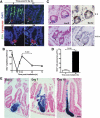
Figure 1
Figure 1
IR induces dose-dependent BM and GI lethality in Lgr5-lacZ transgenic mice. (A) Representative full-transverse section of proximal jejunum from Lgr5-lacZ transgenic mice stained for lacZ. LacZ+ CBCs are visible at the crypt base (blue cells with arrow). (B) Actuarial survival of 8-to 12-week-old Lgr5-lacZ transgenic mice treated with 12 Gy and 15 Gy WBR with/without administration of 5 × 10⁶ syngeneic BM cells 16 hours after irradiation. Actuarial survival was calculated by the Kaplan–Meier method. Number of animals/group is shown in parentheses. (C) Tissue damage in Lgr5-lacZ mice dying after 12 Gy or 15 Gy. H&E-stained sections of femur and proximal jejunum were obtained from animals displaying an agonal-breathing pattern. Murine small intestinal mucosa is well preserved at day 10 after 12 Gy, whereas BM elements appear depleted from the femur cavity. In contrast, mucosa is denuded at day 5 after 15 Gy, with almost no villi/crypts apparent, although BM shows only partial damage. Scale bar, 60 μm.
Figure 2
Figure 2
Close correlation of Lgr5+ stem cell loss to crypt loss. (A) GI damage assessed by the crypt microcolony assay of Withers and Elkind. (B and C) Dose–dependent radiation-induced CBC depletion in Lgr5-lacZ mice. CBC frequency in sectioned LacZ+-stained small intestines was scored at (B) 3.5 or (C) 2 days after irradiation. CBC number/circumference was assessed by counting crypt base lacZ-positive cells. CBC number and crypt survival were quantified from 4 mice/time point, with 3–5 circumferences scored per mouse. Triangles represent the number of crypts or CBCs in individual circumferences. Data (mean ± standard deviation) are from 2 experiments.
Figure 3
Figure 3
Quantitative comparison of CBC-depletion kinetics in small intestines of Lgr5-lacZ transgenic mice treated with 12 Gy and 15 Gy. Shown are CBC kinetic profiles after 12 Gy (right panel) and 15 Gy (left panel). Small intestines were obtained from irradiated Lgr5-lacZ mice and surviving CBCs identified after lacZ-staining as in Figure 1A. CBC survival data (mean ± standard deviation) was quantified from 4 mice per time point, with 3–5 circumferences/mouse. Triangles represent CBC number in individual circumferences. Lower panel: sectioned lacZ-stained small intestines of Lgr5-lacZ mice with/without IR. Scale bar, 20 μm.
Figure 4
Figure 4
CBCs repair DNA damage much faster than other differentiated cells. (A) Confocal images (objective, 20×) of γ-H2AX immunofluorescence staining on small intestinal sections in control and irradiated mice at 6 hours after 12 Gy. Scale bar, 70 μm. (B) Kinetic analysis of γ-H2AX focus resolution in CBCs, TA cells, and villus cells after 12 Gy WBR. Foci/nucleus was determined by counting 3 classes of cells: CBC cells (located between Paneth cells), TA cells (at positions 4–10, with position 4 being directly above Paneth cells), and villus cells (midvillus). Paneth cells were stained for lysozyme (red). Data (mean ± standard error of the mean) are collated from 3 experiments. (C) Representative high-magnification confocal images (objective, 63×) showing γ-H2AX foci on small intestinal sections after 12 Gy. Scale bar, 10 μm. White arrows identify CBCs. *P < .05; **P < .001; each vs CBCs cells.
Figure 5
Figure 5
CBCs manifest rapid formation/resolution of HR and NHEJ foci. (A) Quantification of BRCA1 (left panel) and RAD51 (right panel) foci in CBCs, TA cells, and villus cells as in Figure 4 after 12 Gy WBR. Data (mean ± standard error of the mean) were collated from 3 experiments. Note 36% ± 3% of CBCs and 67% ± 4% of TA cells display RAD51 foci, whereas 100% of both populations display BRCA1 foci. (B) Confocal images (objective, 20×) of BRCA1 immunofluorescence staining on small intestinal sections of control and irradiated mice 1 hour after 12 Gy. Scale bar, 30 μm. (C) Representative high-magnification confocal images (objective, 63×) of (C) BRCA1 and (D) RAD51 foci in crypts after 12 Gy. (E) Quantification of γ-H2AX, BRCA1, and RAD51 foci in CBCs and TA cells at 6 hours after 12 Gy in PUMA knockout mice (right panel). Confocal image (objective, 63×) showing crypt γ-H2AX foci (left panel). Data (mean ± standard deviation) were scored in 100 cells, pooled from 3 independent mice. **P < .001 CBCs vs TA cells. (F) DNA-PKcs foci quantification (left panel) and confocal images (right panel, crypts only) at 30 minutes to 24 hours after 12 Gy. Data (mean ± standard error of the mean) were collated from 3 experiments. Note that at 6, 12, and 24 hours, P < .005 CBCs vs TA cells. Scale bars, 10 μm. White arrows identify CBCs.
Figure 6
Figure 6
Kinetic analysis of CBC cell-cycle arrest and re-initiation after irradiation. (A) Kinetics of cell proliferation after 12 Gy in intestinal sections of 8-week-old Lgr5-lacZ mice pulsed with EdU for 4 hours before death. CBCs (white arrows) are interspersed between Paneth cells stained red for lysozyme. (B) Frequency of EdU-positive crypts/circumference in small intestines of Lgr5-lacZ mice after 12 Gy. Data (mean ± standard error of the mean) were compiled from 3 experiments. (C) LacZ (blue) and BrdU (brown) double staining of consecutive intestinal sections from Lgr5-lacZ mice with/without 12 Gy at 18 hours. Black arrows highlight double-positive CBCs. Scale bar, 10 μm. (D) Percentage of BrdU-labeled Lgr5+ small intestinal cells before and at 18 hours after 12 Gy. Two hundred crypts were counted per mouse with 5 mice/group. Bars represent mean ± standard error of the mean of 3 experiments. (E) Lineage tracing in small intestines of irradiated Lgr5-EGFP-ires-CreERT2/Rosa26-lacZ mice at days 5, 7, and 10 after 12 Gy. Adult mice were injected intraperitoneally with a single tamoxifen dose immediately after radiation. Scale bar, 30 μm.
Figure 7
Figure 7
Kinetic evaluation of apoptotic cell death in CBCs and TA cells. Lgr5-lacZ mice were irradiated with 12 Gy WBR. Apoptotic death of CBCs (black arrows) and TA cells (white arrows) was evaluated by staining for active caspase-3 fragments as described in the Materials and Methods section. CBC cells are located between Paneth cells, although approximately 10% of Lgr5+ CBCs may occur at the +4 position, with position 4 being directly above the most distal Paneth cell. TA cells are located at positions 4–10. Paneth cells are identified by the presence of granules, readily observed under brightfield microscopy. Although Paneth cell degranulation may occur after irradiation, this did not occur to an extent that altered Paneth cell detection in our studies. A minimum of 300 cells were scored for apoptosis per time point. Data (mean ± standard deviation) were collated from 4 mice. Scale bar, 10 μm.

Comment in

References

    1. Mohrin M, Bourke E, Alexander D, et al. Hematopoietic stem cell quiescence promotes error-prone DNA repair and mutagenesis. Cell Stem Cell. 2010;7:174–185. - PMC - PubMed
    1. Sotiropoulou PA, Candi A, Mascre G, et al. Bcl-2 and accelerated DNA repair mediates resistance of hair follicle bulge stem cells to DNA-damage-induced cell death. Nat Cell Biol. 2010;12:572–582. - PubMed
    1. Tichy ED, Pillai R, Deng L, et al. Mouse embryonic stem cells, but not somatic cells, predominantly use homologous recombination to repair double-strand DNA breaks. Stem Cells Dev. 2010;19:1699–1711. - PMC - PubMed
    1. Potten CS, Gandara R, Mahida YR, et al. The stem cells of small intestinal crypts: where are they? Cell Prolif. 2009;42:731–750. - PMC - PubMed
    1. Potten CS. Radiation, the ideal cytotoxic agent for studying the cell biology of tissues such as the small intestine. Radiat Res. 2004;161:123–136. - PubMed

Publication types