. 2012 Oct 15;303(8):L642-60.
doi: 10.1152/ajplung.00406.2011. Epub 2012 Jul 27.

Inhibition of allergic inflammation by supplementation with 5-hydroxytryptophan

Affiliations

Inhibition of allergic inflammation by supplementation with 5-hydroxytryptophan

Hiam Abdala-Valencia et al. Am J Physiol Lung Cell Mol Physiol. .

Abstract

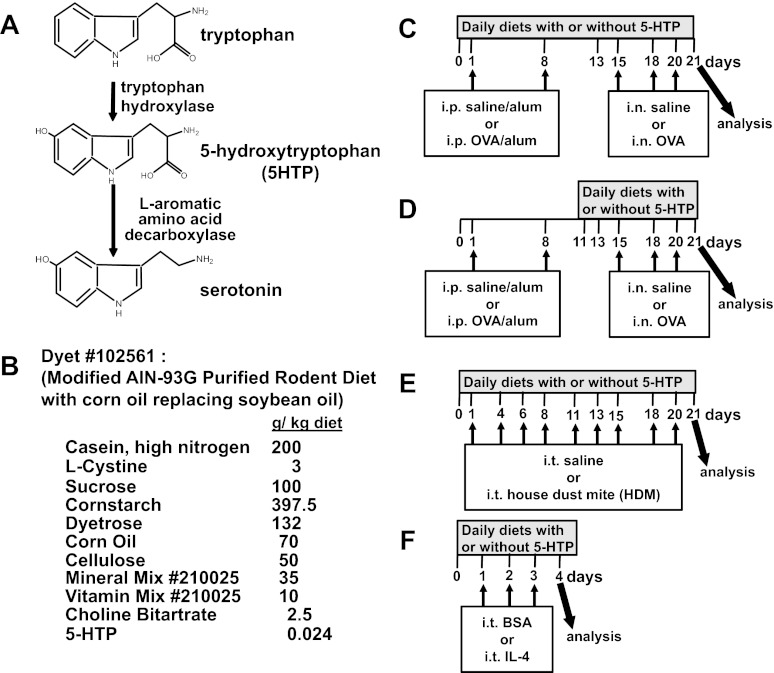
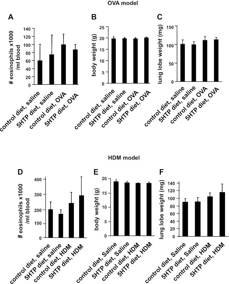
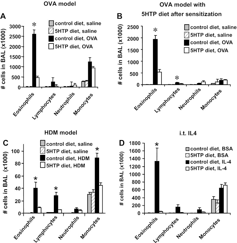
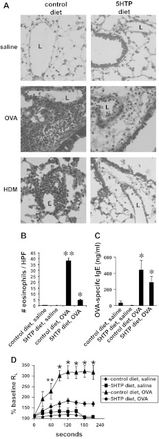
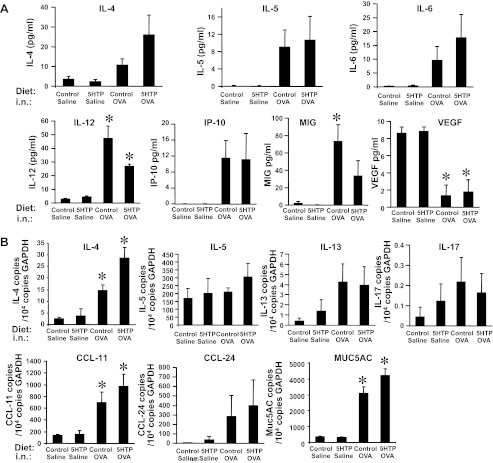
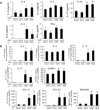
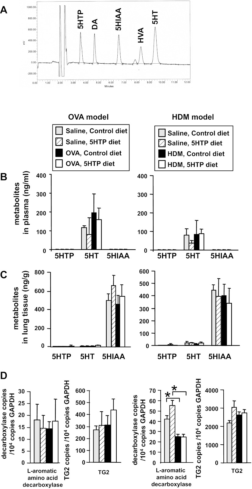
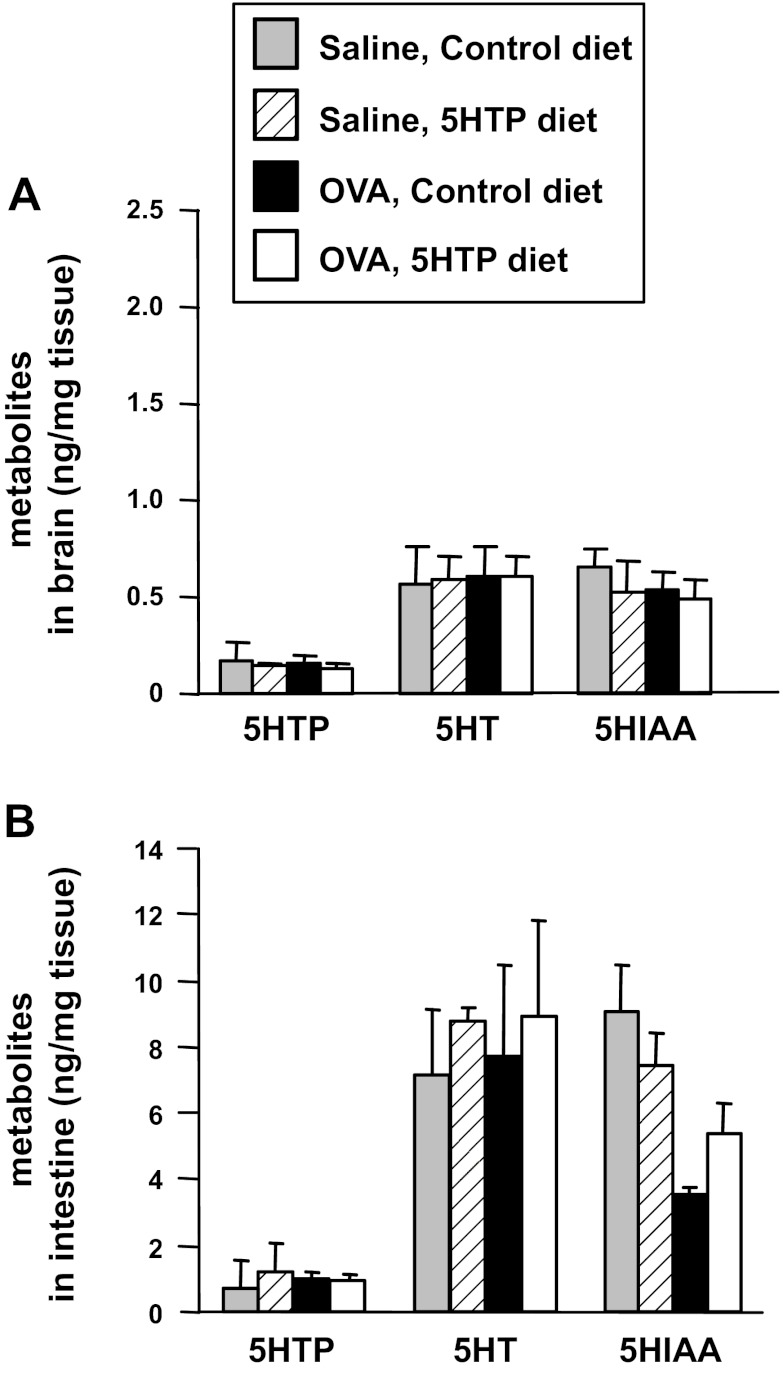
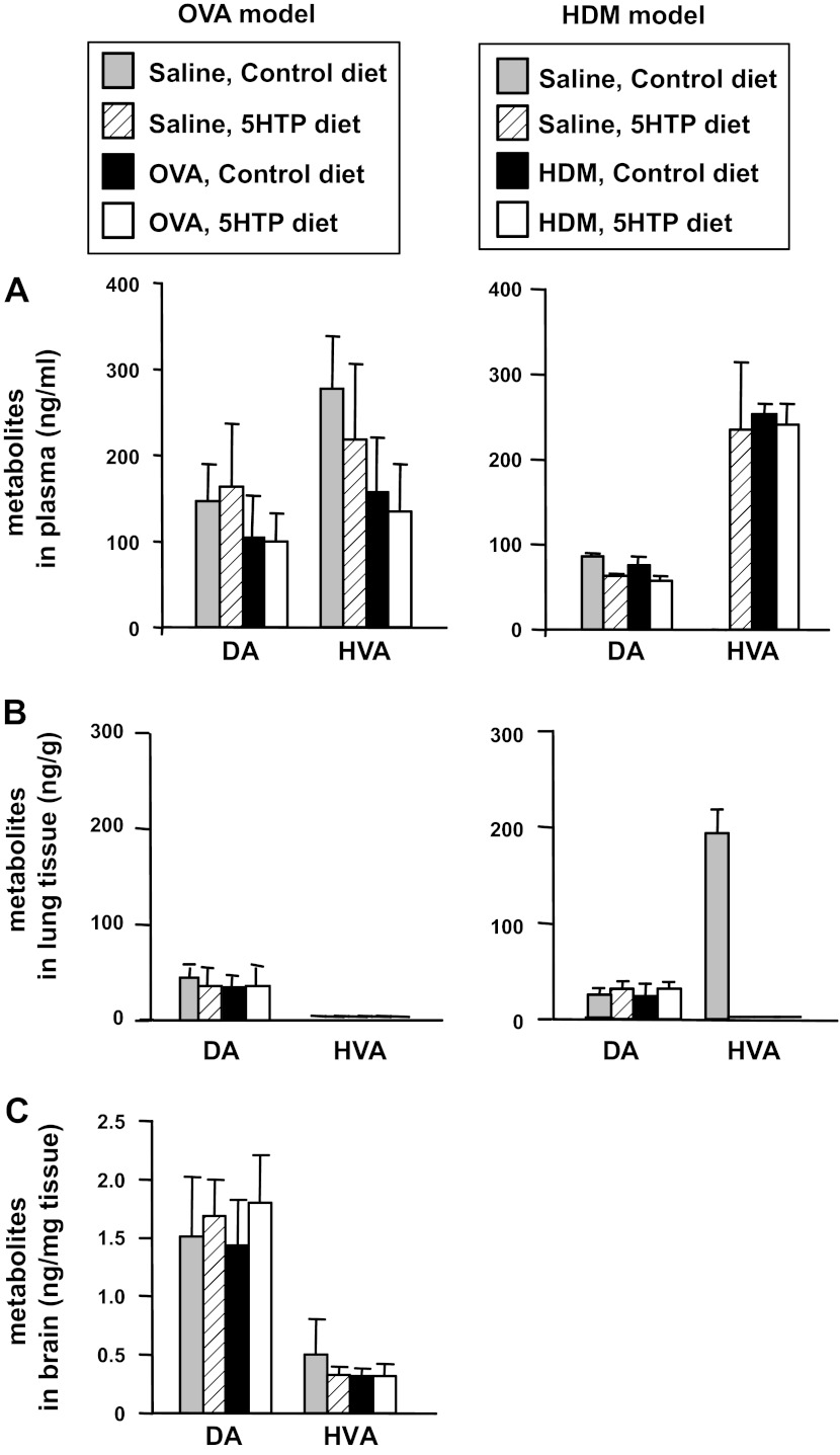
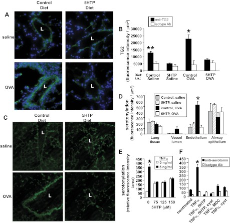
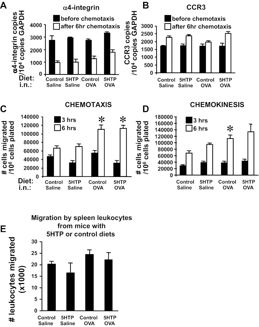
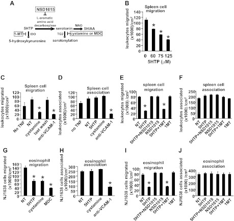
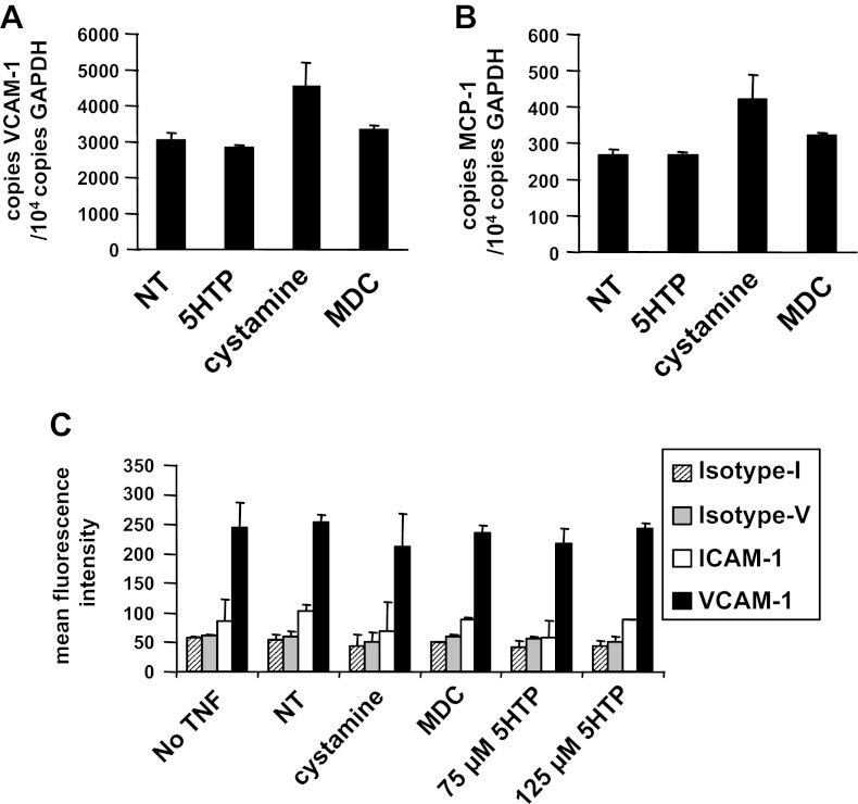
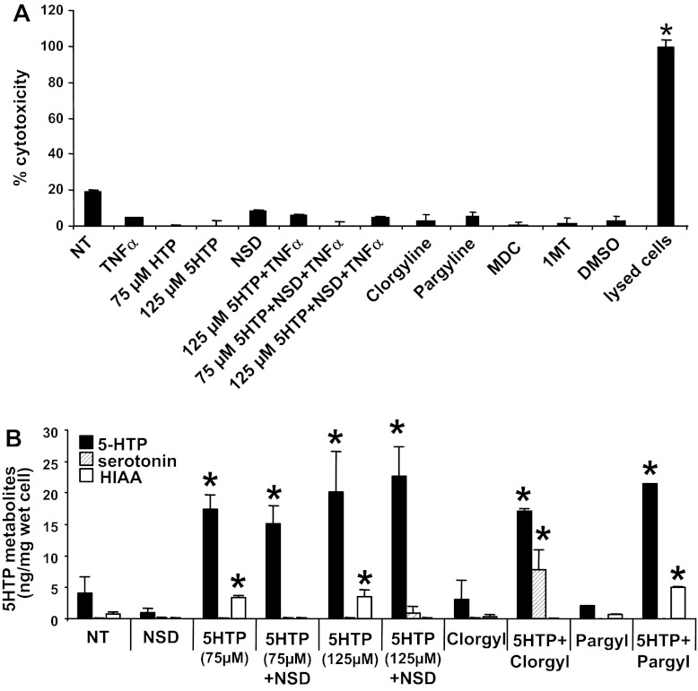
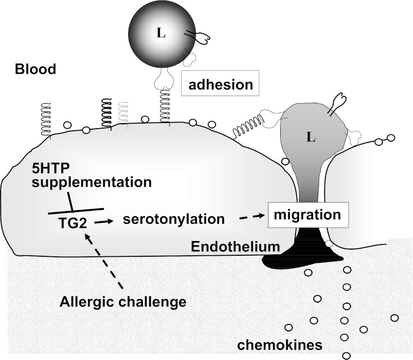
Clinical reports indicate that patients with allergy/asthma commonly have associated symptoms of anxiety/depression. Anxiety/depression can be reduced by 5-hydroxytryptophan (5-HTP) supplementation. However, it is not known whether 5-HTP reduces allergic inflammation. Therefore, we determined whether 5-HTP supplementation reduces allergic inflammation. We also determined whether 5-HTP decreases passage of leukocytes through the endothelial barrier by regulating endothelial cell function. For these studies, C57BL/6 mice were supplemented with 5-HTP, treated with ovalbumin fraction V (OVA), house dust mite (HDM) extract, or IL-4, and examined for allergic lung inflammation and OVA-induced airway responsiveness. To determine whether 5-HTP reduces leukocyte or eosinophil transendothelial migration, endothelial cells were pretreated with 5-HTP, washed and then used in an in vitro transendothelial migration assay under laminar flow. Interestingly, 5-HTP reduced allergic lung inflammation by 70-90% and reduced antigen-induced airway responsiveness without affecting body weight, blood eosinophils, cytokines, or chemokines. 5-HTP reduced allergen-induced transglutaminase 2 (TG2) expression and serotonylation (serotonin conjugation to proteins) in lung endothelial cells. Consistent with the regulation of endothelial serotonylation in vivo, in vitro pretreatment of endothelial cells with 5-HTP reduced TNF-α-induced endothelial cell serotonylation and reduced leukocyte transendothelial migration. Furthermore, eosinophil and leukocyte transendothelial migration was reduced by inhibitors of transglutaminase and by inhibition of endothelial cell serotonin synthesis, suggesting that endothelial cell serotonylation is key for leukocyte transendothelial migration. In summary, 5-HTP supplementation inhibits endothelial serotonylation, leukocyte recruitment, and allergic inflammation. These data identify novel potential targets for intervention in allergy/asthma.


Figures

Fig. 1.
Fig. 1.
5-Hydroxytryptophan (5-HTP) supplementation. A: serotonin synthesis. B: purified 5-HTP from Sigma was used to supplement the diets by Dyets. Consumption of this diet (0.024 g 5-HTP/kg diet) at 4 g diet/day/20 g mouse is equivalent to 200 mg 5-HTP/100 pound person/day. This calculation is as follows: 200 mg 5-HTP/100 pound person/day = {[(0.024 g 5-HTP × 1,000 mg/g)/1,000 g diet] × 4 g diet/mouse/day}/[20 g mouse × (1 pound/453 g)]. The control diet was the same except without 5-HTP. We confirmed the concentration of 5-HTP in the diet as 0.024 g 5-HTP/kg diet by HPLC/electrochemical detector (ECD). C: timeline for 5-HTP supplementation during ovalbumin fraction V (OVA) sensitization and OVA challenge treatments. D: timeline for 5-HTP supplementation after OVA sensitization. E: timeline for 5-HTP supplementation during treatments with house dust mite (HDM) extract from Dermatophagoides pteronyssius. F: timeline for 5-HTP supplementation during intratracheal IL-4 treatments. On day of analysis in C–F, the bronchoalveolar lavage (BAL), perfused lungs, blood, brains, and intestines were collected. i.p., intraperitoneal. i.n., intranasal. i.t., intratracheal. n = 6–10 mice/group.
Fig. 2.
Fig. 2.
5-HTP supplementation did not alter blood eosinophils, body weight, or lung weight. Mice were supplemented with 5-HTP and received the antigens OVA (A–C) or HDM (D–F) as in the timeline Fig. 1, C and E. The mice were weighed, and blood eosinophils were counted. A and D: blood eosinophils. B and E: mouse body weight. C and F: mouse lung lobe weight. n = 8–10 mice/group. The groups are not statistically different.
Fig. 3.
Fig. 3.
5-HTP supplementation reduced the number of leukocytes in the BAL. A: mice were supplemented with 5-HTP and received OVA as in the timeline Fig. 1C. n = 8–10 mice/group. B: mice were supplemented with 5-HTP after OVA sensitization as in the timeline in Fig. 1D. n = 6–8 mice/group. C: mice were supplemented with 5-HTP and received HDM as in the timeline Fig. 1E. n = 8–10 mice/group. D: mice received intratracheal (i.t.) administration of 4 μg IL-4/mouse/day or the protein control BSA grade VI as in the timeline in Fig. 1F. n = 6 mice/group. The BAL cells were collected and cytospun and then neutrophils, eosinophils, monocytes, and lymphocytes were counted by standard morphological criteria. *P < 0.05 compared with the other groups.
Fig. 4.
Fig. 4.
5-HTP supplementation reduced allergen-induced lung tissue inflammation and lung responsiveness. A: frozen lung tissue sections from the mice in Fig. 3, A and C, were stained with hematoxylin and eosin. Shown are representative micrographs of perivascular regions in lung tissue. n = 8–10 mice/group. L, vessel lumen. B: number of antimajor basic protein (MBP)-labeled lung perivascular eosinophils per high-powered field (HPF) from mice in Fig. 3A. C: 5-HTP diets did not alter plasma OVA-specific IgE from mice in Fig. 3A as measured by ELISA. n = 8–10 mice/group. D: to examine OVA-induced lung resistance, OVA-treated mice were retro-orbitally challenged with 500 μg OVA VI in 50 μl saline or saline alone (29). Exposure to mechanical ventilation and measurements of lung mechanics were performed using a flexiVent mouse ventilator. Presented are the percentages of baseline lung resistance (RL). **P < 0.05 compared with saline groups. *P < 0.05 compared with all other groups.
Fig. 5.
Fig. 5.
For OVA-challenged mice, 5-HTP supplementation did not alter expression of lung cytokines, chemokines, growth factors, or mucin. The lungs and BAL were collected from the OVA-challenged mice in Fig. 3A. A: BAL supernatants from lungs in Fig. 3A were tested for levels of cytokines using the Luminex 20plex cytokine kit. Not detected were IFN-γ, IL-1α, IL-1β, IL-2, KC, monocyte chemoattractant protein (MCP)-1, and TNF-α; IL-17 was very low (data not shown). B: lung tissue from mice in Fig. 3A was placed in RNAlater and then examined by quantitative PCR for IL-4, IL-5, IL-13, IL-17, CCL11, CCL24, and MUC5AC expression. n = 8–10 mice/group. *P < 0.05 compared with the saline groups. There is not a significant effect of 5-HTP supplementation of OVA-treated mice compared with control diet OVA-treated mice.
Fig. 6.
Fig. 6.
For the HDM-challenged mice, 5-HTP supplementation did not alter lung cytokines, chemokines, mucin, or adhesion molecules. The lungs and BAL were collected from the HDM-challenged mice in Fig. 3C. A: BAL supernatants were tested for levels of cytokines using the Luminex 6plex cytokine kit. B: lung tissue was placed in RNAlater and then examined by quantitative PCR for IL-4, IL-5, IL-13, IL-17, VCAM-1, CCL11, CCL24, and MUC5AC. n = 8–10 mice/group. *P < 0.05 compared with the saline groups. There is not a significant effect of 5-HTP supplementation of HDM-treated mice compared with control diet HDM-treated mice.
Fig. 7.
Fig. 7.
5-HTP supplementation did not alter 5-HTP or its metabolites in plasma or perfused whole lung tissue. A: HPLC/ECD profile of standards demonstrating separation of 5-HTP and its metabolites serotonin (5-HT) and 5-hydroxyindoleacetic acid (HIAA) as well as dopamine (DA) and its metabolite homovanillic acid (HVA). The solvent peaks come off the column before the metabolites (unlabeled peaks). Peaks are labeled with the metabolite. From mice in Fig. 3, A and C, plasma (B) and lungs (C), which had been perfused free of blood, were examined for 5-HTP, 5-HT, and HIAA by HPLC with electrochemical detection. D: lung tissue was placed in RNAlater and then examined by quantitative PCR for l-aromatic amino acid decarboxylase and transglutaminase (TG)2 in total lung tissue. n = 8–10 mice/group. *P < 0.05 for the indicated groups.
Fig. 8.
Fig. 8.
5-HTP supplementation did not alter 5-HTP or its metabolites in brain or intestine. Brain and intestines were collected from OVA-challenged mice from Fig. 3A and frozen. Brains (A) and intestines (B) were examined for 5-HTP, 5-HT, and HIAA by HPLC/ECD. n = 8–10 mice/group. There is not a significant difference among the treatment groups.
Fig. 9.
Fig. 9.
5-HTP supplementation did not alter dopamine or its metabolite HVA in plasma, perfused whole lung tissue, or brain. From mice in Fig. 3, A and C, plasma (A), lungs that had been perfused free of blood (B), and brains (C) were examined for DA and HVA by HPLC/ECD. DA and HVA were not detected in intestines (data not shown). n = 8–10 mice/group. There is not a significant difference among the treatment groups.
Fig. 10.
Fig. 10.
5-HTP supplementation reduced OVA-induced lung vascular TG2 expression and serotonylation in vivo and reduced TNF-α-stimulated endothelial cell serotonylation in vitro. A–D: tissue sections from perfused lungs of mice from Fig. 3A were labeled with anti-TG2 or antiserotonin antibodies and examined by fluorescence microscopy. Labeling with isotype control antibodies was negative such that the fluorescence was lower than the intensity in the vessel lumen of the saline control groups (data not shown). A: anti-TG2 immunofluorescence labeling (green) and DAPI (blue). Representative micrographs with vessels. B: fluorescence intensity of TG2/μm2 of vessel endothelium. C: antiserotonin immunofluorescence labeling. Representative micrographs with vessels. D: fluorescence intensity of serotonylation/μm2 of vessel endothelium, vessel lumen, airway epithelium, or lung tissue. B and D: for each cell type, data from 3 sections per mouse were collected and averaged to obtain an average for each mouse. Then the data in the graphs are from the average ± the SE of 8 mice/group. E–F: in vitro, monolayers of endothelial cells were stimulated with TNF-α in the presence or absence of 125 μM 5-HTP, and/or the TG inhibitors cystamine (Cyst, 100 μM) or dansylcadaverine (MDC, 200 μM). n = 3–5 for in vitro serotonylation. The inhibitors did not affect cell viability (shown in Fig. 14). L, vessel lumen. *P < 0.05 compared with the other treatment groups. **P < 0.05 compared with the other treatment groups.
Fig. 11.
Fig. 11.
For OVA-challenged mice, 5-HTP supplementation of leukocytes did not alter expression of leukocyte α4-integrin, the chemokine receptor CCR3, chemotaxis, chemokinesis, or transendothelial migration. A and B: spleen leukocytes from mice in Fig. 3B were placed in RNAlater before and after the chemotaxis in C. These cells were examined by quantitative PCR for α4-integrin (A) and CCR3 expression (B). C and D: chemotaxis and chemokinesis of spleen cells isolated from mice in Fig. 3B. The assays were performed with 50 ng CCL-11/ml for 3 or 6 h. E: transendothelial migration by spleen leukocytes from mice supplemented with 5-HTP as in Fig. 3B. Leukocytes were added to confluent monolayers of endothelial cells in 8-μm-pore Transwells overnight. The migrated leukocytes in the bottom chamber were counted with a hemocytometer. i.n., intranasal. n = 8–10 mice/group. *P < 0.05 compared with the saline groups. There is not a significant effect of 5-HTP supplementation of OVA-treated mice compared with control diet OVA-treated mice.
Fig. 12.
Fig. 12.
5-HTP pretreatment of endothelial cells in vitro reduced leukocyte transendothelial migration. A: diagram of 5-HTP metabolism to HIAA or serotonylation. Also indicated is the potential 5-HTP metabolism to 5-hydroxykynurenine; it is reported that 5-HTP application to intestines ex vivo generates 5-hydroxykynurenine (46). In boxes are the inhibitors. B–J: endothelial cell monolayers were pretreated with 5 ng/ml TNF-α overnight except where indicated as “no TNF”. TNF-α-stimulated endothelial cell monolayers on slide flasks were pretreated overnight as indicated with 5-HTP (75–125 μM), the TG2 inhibitors cystamine (100 μM) and dansylcadaverine (MDC, 200 μM), the aromatic amino acid decarboxylase inhibitor NSD1015 (30 μM), or the indolamine dioxygenase (IDO) inhibitor 1-MT (100 μM 1-l-methyl-tryptophan + 100 μM 1-d-methyl-tryptophan) and then washed before addition of BALB/c mouse spleen leukocytes (>90% lymphocytes) (B–F) or NJ1638 mouse eosinophils (>85% eosinophils) (G–J) for the migration and adhesion assays in a parallel plate flow chamber. The above inhibitors did not affect endothelial cell viability (data not shown) or cell cytotoxicity (Fig. 14A). Where indicated, anti-VCAM-1 antibodies (54 μg Ab/800 μl/slide flask) were added to the endothelial cells 15 min before addition of leukocytes. B–J: comparisons can only be made within an experiment because total migration can vary somewhat among experiments. In C, the “last wash” designates cells that were treated with the 5th wash from cystamine-pretreated endothelial cells, indicating that the cells were sufficiently washed to remove effective concentrations of the inhibitor. NT, nontreated. MAO, monoamine oxygenase. n = 3–5. *P < 0.05 compared with nontreated controls.
Fig. 13.
Fig. 13.
5-HTP pretreatment of endothelial cells did not alter endothelial cell expression of VCAM-1 or MCP-1. A and B: TNF-α-treated endothelial cells were incubated overnight with 125 μM 5-HTP, 100 μM cystamine, or 200 μM MDC as in Fig. 12, and the cells were placed in RNAlater; the samples were then examined by quantitative PCR for expression of the adhesion molecule VCAM-1 (A) or the chemokine MCP-1 (B). C: endothelial cells were treated as indicated, immunolabeled with anti-ICAM-1 or an isotype antibody control for the ICAM-1 antibody (isotype-I) or immunolabeled with anti-VCAM-1 or an isotype antibody control for the VCAM-1 antibody (isotype-V), and examined by flow cytometry. The endothelial cell lines expressed VCAM-1 but not ICAM-1 as previously reported (24, 25). n = 2 experiments.
Fig. 14.
Fig. 14.
Endothelial cell metabolism of 5-HTP to serotonin or HIAA was blocked by the inhibitors without inducing endothelial cell cytotoxicity. A: inhibitors in Figs. 10, 12, and 13 did not induce cell cytotoxicity. The endothelial cell monolayers were treated for 15 min with the following inhibitors used in Figs. 10, 12, and 13: 5-HTP (75 or 125 μM), the aromatic amino acid decarboxylase inhibitor NSD1015 (NSD, 30 μM), the monoamine oxygenase inhibitors clorgyline (Clorgyl, 1 μM) or pargyline (Pargyl, 100 μM), the TG inhibitor dansylcadaverine (MDC, 100 μM), the IDO inhibitor 1-MT (100 μM 1-l-methyl-tryptophan + 100 μM 1-d-methyl-tryptophan), or the solvent control 0.01% DMSO. Then the cells were stimulated with 5 ng/ml TNF-α. After overnight culture, the endothelial cells were examined for cell cytotoxicity with the Vybrant Cytotoxicity assay. A set of cells were lysed as a positive control for cytotoxicity. B: endothelial cells were pretreated with NSD1015 (NSD, 30 μM), 5-HTP (75 or 125 μM), clorgyline (1 μM), or pargyline (Pargyl, 100 μM) where indicated and then stimulated with 5 ng/ml TNF-α. After overnight culture, the endothelial cells were examined for 5-HTP, serotonin (5-HT), and HIAA by HPLC/ECD. n = 3. *P < 0.05 indicates a significant increase compared with the nontreated (NT) group.
Fig. 15.
Fig. 15.
Overview of 5-HTP inhibition of endothelial cell function during leukocyte transendothelial migration. During allergic inflammation, tissue-derived cytokines activate endothelial cells to increase expression of TG2, adhesion molecules, and chemokines. Furthermore, endothelial cell TG2-mediated serotonylation increases leukocyte transendothelial migration in response to chemokines. 5-HTP supplementation blocks allergen-induced increases in TG2 expression, TG2-mediated serotonylation, and leukocyte transendothelial migration. L, leukocyte.

References

    1. Abdala-Valencia H, Cook-Mills JM. VCAM-1 signals activate endothelial cell protein kinase cα via oxidation. J Immunol 177: 6379–6387, 2006 - PMC - PubMed
    1. Abdala-Valencia H, Earwood J, Bansal S, Jansen M, Babcock G, Garvy B, Wills-Karp M, Cook-Mills JM. Nonhematopoietic NADPH oxidase regulation of lung eosinophilia and airway hyperresponsiveness in experimentally induced asthma. Am J Physiol Lung Cell Mol Physiol 292: L1111–L1125, 2007 - PMC - PubMed
    1. Abdouh M, Albert PR, Drobetsky E, Filep JG, Kouassi E. 5-HT1A-mediated promotion of mitogen-activated T and B cell survival and proliferation is associated with increased translocation of NF-kappaB to the nucleus. Brain Behav Immun 18: 24–34, 2004 - PubMed
    1. Abdouh M, Storring JM, Riad M, Paquette Y, Albert PR, Drobetsky E, Kouassi E. Transcriptional mechanisms for induction of 5-HT1A receptor mRNA and protein in activated B and T lymphocytes. J Biol Chem 276: 4382–4388, 2001 - PubMed
    1. Alberghina D, Amorini AM, Lazzarino G. Modulation of peripheral markers of the serotoninergic system in healthy horses. Res Vet Sci 90: 392–395, 2011 - PubMed

Publication types

MeSH terms