Case Reports
. 2012 Sep;44(9):1040-5.
doi: 10.1038/ng.2361. Epub 2012 Jul 29.

NMNAT1 mutations cause Leber congenital amaurosis

Affiliations
Case Reports

NMNAT1 mutations cause Leber congenital amaurosis

Marni J Falk et al. Nat Genet. 2012 Sep.

Abstract

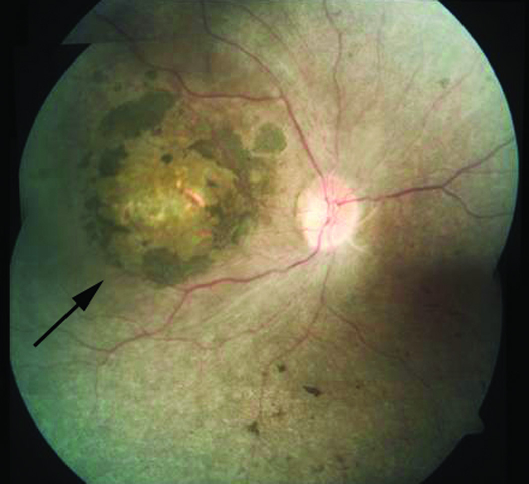
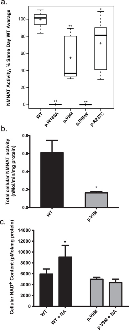
Leber congenital amaurosis (LCA) is an infantile-onset form of inherited retinal degeneration characterized by severe vision loss(1,2). Two-thirds of LCA cases are caused by mutations in 17 known disease-associated genes(3) (Retinal Information Network (RetNet)). Using exome sequencing we identified a homozygous missense mutation (c.25G>A, p.Val9Met) in NMNAT1 that is likely to be disease causing in two siblings of a consanguineous Pakistani kindred affected by LCA. This mutation segregated with disease in the kindred, including in three other children with LCA. NMNAT1 resides in the previously identified LCA9 locus and encodes the nuclear isoform of nicotinamide mononucleotide adenylyltransferase, a rate-limiting enzyme in nicotinamide adenine dinucleotide (NAD(+)) biosynthesis(4,5). Functional studies showed that the p.Val9Met alteration decreased NMNAT1 enzyme activity. Sequencing NMNAT1 in 284 unrelated families with LCA identified 14 rare mutations in 13 additional affected individuals. These results are the first to link an NMNAT isoform to disease in humans and indicate that NMNAT1 mutations cause LCA.


Figures

Figure 1
Figure 1
Pedigrees of three LCA kindreds evaluated at The Children’s Hospital of Philadelphia in whom mutations were identified in NMNAT1. (a) Family 047. Consanguineous Pakistani kindred in which homozygous NMNAT1 mutations were identified in five children with LCA by whole exome sequencing with Sanger sequencing validation. M1 = NMNAT1 mutation (c.25G>A, p.Val9Met). M2 = GJB2 mutation (c.71G>A , p.Trp24*). A representative sequence trace for the M1 mutation is shown. Exome sequencing confirmed the presence of the p.Trp24* homozygous mutation in GJB2 in the two children (IV-1 and IV-2) who were affected with sensorineural hearing loss. Sanger sequencing verified that this mutation also segregated with the hearing loss phenotype in their larger kindred. Subject IV-2 had no identifiable pathogenic mutations within any of the known mucolipidosis disease genes. (b) The members of family 047 were genotyped by PCR amplification of exon 2 of NMNAT1, followed by digestion of the PCR products with AcuI to distinguish the wild-type from the mutant sequence. The genotyping data illustrate the segregation of the mutant NMNAT1 (M1) allele only in the five children with LCA in generation IV, whereas the four unaffected parents of these children in generation III carry both the mutant and wild-type alleles, and their three children with normal vision harbor only the wild-type allele. Individuals III-4, III-5, IV-1, IV-2 and IV-3 were clinically evaluated by M.J.F. and E.A.P.; individuals III-3, III-6 and V-4 to IV-7 were not clinically evaluated by the authors. (c) Family 007. A single LCA proband was identified by Sanger sequencing of NMNAT1 to harbor compound heterozygous mutations c.196C>T (p.Arg66Trp) and c.709C>T (p. Arg237Cys). (d) Family 053. A single LCA proband was identified by Sanger sequencing of NMNAT1 to harbor compound heterozygous mutations c.205A>G (p.Met69Val) and c.769G>A (p.Glu257Lys). The ‘+’ or ‘M’ below each symbol represents a wild-type or specific mutant allele. Squares and circles indicate male or female, respectively, and numbers within symbols indicate multiple offspring of a given gender. Slashes depict deceased individuals. Colors within symbols indicate affected individuals, with phenotypes defined in the pedigree key.
Figure 2
Figure 2
Retinal image from individual with LCA due to mutations in NMNAT1. Composite fundus image of the right eye of subject II-1, LVPEI family LCA-100, showing pallor of the optic disc, attenuation of the retinal blood vessels, pigment disruption and atrophic changes in the macula (arrow), and scattered pigment clumping in the peripheral retina. The optic disc is approximately 1.75 mm in diameter.
Figure 3
Figure 3
NMNAT1 enzyme activity and cellular NAD+ content. (a) The NAD+ biosynthetic activity of wild-type, p.Val9Met, p.Arg66Trp, p.Arg237Cys and p.Trp169Ala purified recombinant NMNAT1 proteins was measured as described. Boxplots display activity measurements of independently generated and measured replicate protein preparations. The length of the box represents 25th to 75th inter-quartile range, the interior horizontal line represents the median, the interior cross represents the mean, and vertical lines issuing from the box extend to the minimum and maximum values of the analysis variable. The p.Trp169Ala mutant had complete loss of NMNAT1 enzyme activity (n = 6; P = 0.0014), as was previously reported. The p.Val9Met mutant protein NMNAT1 enzyme activity was significantly reduced at 37% of wild-type control (n = 7; P = 0.0015). The p.Arg237Cys mutant protein showed an average of 81% of wild-type NMNAT1 activity (n = 6; P = 0.034). The p.Arg66Trp mutant protein showed complete loss of enzyme activity (n = 6; P = 0.0014), although we were not able to achieve effective purification of the Flag-tagged version of the p.Arg66Trp mutant protein (Supplementary Fig. 6) despite its clearly normal expression and nuclear localization in CHO and IMCD3 cells (Supplementary Fig. 5). Further experiments will be needed to determine if the Flag-tagged p.Arg66Trp protein is unstable to purification. *P < 0.05 and **P < 0.01 determined by non-parametric Wilcoxon rank sum test. (b) Total cellular NMNAT enzyme activity was measured in whole cell extracts of fibroblasts from a healthy control and the LCA proband from Family 047 (subject IV-1) who was homozygous for the p.Val9Met NMNAT1 variant. The p.Val9Met mutant cells had 27% of total cellular NMNAT NAD+ synthetic activity relative to control cells (two-tailed t test P = 0.016; n = 6 for wild-type, n = 5 for LCA proband cells). (c) Cellular NAD+ levels. Total cellular NAD+ content was quantified by HPLC in control and LCA proband IV-1 (Fig. 1a) fibroblasts at baseline and following 10 mM nicotinic acid treatment for 24 hours. NAD+ content in fibroblast cells from the LCA proband (p.Val9Met) was decreased by 16% relative to those from a wild-type control (P = 0.067). Nicotinic acid treatment significantly increased NAD+ content in control cells (P < 0.05) but had no effect on NAD+ content in the proband’s cells (P > 0.05). n = 7 for both cell lines without treatment, n = 6 for control cells treated with nicotinic acid, and n = 4 for IV-1 cells treated with nicotinic acid. For b and c, bars indicate mean and standard error. *P < 0.05.

Comment in

Similar articles

Cited by

References

    1. Weleber RG. Infantile and childhood retinal blindness: a molecular perspective. Ophthalmic Genet. 2002:71–97. - PubMed
    1. Michaelides M, Hardcastle AJ, Hunt DM, Moore AT. Progressive cone and cone-rod dystrophies: phenotypes and underlying molecular genetic basis. Survey of Ophthalmology. 2006;51:232–258. - PubMed
    1. den Hollander AI, Black A, Bennett J, Cremers FP. Lighting a candle in the dark: advances in genetics and gene therapy of recessive retinal dystrophies. J. Clin. Invest. 2010;120:3042–3053. - PMC - PubMed
    1. Keen TJ, et al. Identification of a locus (LCA9) for Leber's congenital amaurosis on chromosome 1p36. Eur. J. Hum. Genet. 2003;11:420–423. - PubMed
    1. Lau C, Niere M, Ziegler M. The NMN/NaMN adenylyltransferase (NMNAT) protein family. Front. Biosci. 2009;14:410–431. - PubMed

Publication types

MeSH terms

Substances