. 2012 Nov;47(5):660-8.
doi: 10.1165/rcmb.2012-0047OC. Epub 2012 Jul 27.

Regulation of Rela/p65 and endothelial cell inflammation by proline-rich tyrosine kinase 2

Affiliations

Regulation of Rela/p65 and endothelial cell inflammation by proline-rich tyrosine kinase 2

Kaiser M Bijli et al. Am J Respir Cell Mol Biol. 2012 Nov.

Abstract

We investigated the role of proline-rich tyrosine kinase 2 (Pyk2) in the mechanism of NF-κB activation and endothelial cell (EC) inflammation induced by thrombin, a procoagulant serine protease released in high amounts during sepsis and other inflammatory conditions. Stimulation of ECs with thrombin resulted in a time-dependent activation of Pyk2. RNA interference knockdown of Pyk2 attenuated thrombin-induced activity of NF-κB and expression of its target genes, vascular cell adhesion molecule-1 and monocyte chemoattractant protein-1. Pyk2 knockdown impaired thrombin-induced activation of IκB kinase (IKK) and phosphorylation (Ser32 and Ser36) of IkappaBα, but, surprisingly, failed to prevent IκBα degradation. However, depletion of IKKα or IKKβ was effective in inhibiting IκBα phosphorylation/degradation, as expected. Intriguingly, Pyk2 knockdown impaired nuclear translocation and DNA binding of RelA/p65, despite the inability to prevent IκBα degradation. In addition, Pyk2 knockdown was associated with inhibition of RelA/p65 phosphorylation at Ser536, which is important for transcriptional activity of RelA/p65. Depletion of IKKα or IKKβ each impaired RelA/p65 phosphorylation. Taken together, these data identify Pyk2 as a critical regulator of EC inflammation by virtue of engaging IKK to promote the release and the transcriptional capacity of RelA/p65, and, additionally, by its ability to facilitate the nuclear translocation of the released RelA/p65. Thus, specific targeting of Pyk2 may be an effective anti-inflammatory strategy in vascular diseases associated with EC inflammation and intravascular coagulation.


Figures
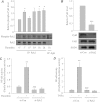
Figure 1.
Figure 1.
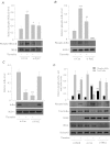
(A) Thrombin induces phosphorylation of proline-rich tyrosine kinase 2 (Pyk2) in endothelial cells (ECs). Confluent human pulmonary artery EC (HPAEC) monolayers were challenged with thrombin (5 U/ml) for the indicated time periods. Total cell lysates were subjected to immunoprecipitation (IP) with an antibody to Pyk2 or IgG, as described in Materials and Methods. The immunoprecipitates were then immunoblotted with an antibody to phosphorylated (Tyr402) form of Pyk2 or an anti-Pyk2 antibody. The bar graph represents the level of Pyk2 phosphorylation induced by thrombin at different time points. The phosphorylated Pyk2 level normalized to the total Pyk2 is expressed relative to untreated control set at 1. Data are means (±SE) (n = 3 for each condition); *P < 0.05 compared with thrombin-untreated control. (B) RNA interference knockdown of Pyk2. Cells were transfected with Pyk2 short interfering RNA (si-Pyk2) or with control siRNA (si-Con) for 24–36 hours, as described in Materials and Methods. Total cell lysates were immunoblotted with an antibody to Pyk2 or focal adhesion kinase (FAK). Equal protein loading was monitored by immunoblotting the lysates with an antibody to Cu-Zn superoxide dismutase (SOD-1). The bar graph represents the level of Pyk2 in si-Pyk2 transfected cells. Pyk2 level normalized to SOD-1 level is expressed relative to si-Con set at 1. Data are means (±SE) (n = 5 for each condition); ***P < 0.001 compared with cells transfected with si-Con. (C and D) Pyk2 mediates thrombin- and TNF-α–induced NF-κB activity. HPAECs were transfected with si-Pyk2 or si-Con using DharmaFect1. Twelve hours later, cells were again transfected with NF-κB luciferase (LUC) construct using diethylaminoethyl (DEAE)-Dextran, as described in Materials and Methods. After 12 hours, cells were challenged with thrombin (5 U/ml) (C) or TNF-α (100 U/ml) (D) for 6 hours. Cell extracts were prepared and assayed for Firefly and Renilla luciferase activities. Data are means (±SE) (n = 4–6 for each condition). **P < 0.01 compared with untreated controls; ###P < 0.001 compared with thrombin- or TNF-α–stimulated controls.
Figure 2.
Figure 2.
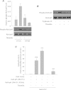
(A and B) Effect of depletion or inhibition of Pyk2 on thrombin-induced vascular cell adhesion molecule (VCAM)-1 expression. HPAECs were transfected with si-Pyk2 or si-Con for 24–36 hours (A) or pretreated with tyrphostin A9 (5 μM) for 1 hour (B). Cells were challenged with thrombin (5 U/ml) for 6 hours. Total cell lysates were immunoblotted with an antibody to VCAM-1. SOD-1 levels were used to monitor loading. The bar graph represents the effect of Pyk2 depletion on VCAM-1 protein expression normalized to SOD-1 level. Data are means (±SE) (n = 3–5 for each condition). ***P < 0.001 compared with untreated controls; ##P < 0.01 compared with thrombin-stimulated controls. (C) Effect of Pyk2 depletion on monocyte chemoattractant protein (MCP)-1. HPAECs were transfected with si-Pyk2 or si-Con for 24–36 hours and then challenged with thrombin (5 U/ml) for 6 hours. Total cell lysates were used to determine MCP-1 levels by ELISA, as described in Materials and Methods. Data are means (±SE) (n = 5–11 for each condition). ***P < 0.001 compared with untreated controls; ###P < 0.001 compared with thrombin-stimulated controls.
Figure 3.
Figure 3.
Effect of Pyk2 depletion on thrombin-induced IKKβ phosphorylation, and IκBα phosphorylation and degradation. HPAECs were transfected with si-Pyk2 or si-Con for 24–36 hours and then challenged with thrombin (5 U/ml) for 1 hour. (A) Total cell lysates were immunoblotted with an anti-phospho IKKα (Ser180)/IKKβ (Ser181) antibody. Total IKKβ levels were used to monitor loading. The bar graph represents the effect of Pyk2 depletion on thrombin-induced phosphorylation of IKKα/β normalized to total IKKβ levels. Data are means (±SE) (n = 3 for each condition). **P < 0.01 compared with untreated controls; #P < 0.05 compared with thrombin-stimulated controls. (B) Lysates were immunoblotted with an anti-phospho IκBα (Ser32 and Ser36) antibody. SOD-1 levels were used to monitor loading. The bar graph represents the effect of Pyk2 depletion on thrombin-induced phosphorylation of IκBα normalized to SOD-1 levels. Data are means (±SE) (n = 3–4 for each condition). ***P < 0.001 compared with untreated controls; ##P < 0.01 compared with thrombin-stimulated controls. (C) Cytoplasmic extracts were immunoblotted with an antibody to IκBα. SOD-1 levels were used to monitor loading. The bar graph represents the effect of Pyk2 depletion on thrombin-induced degradation of IκBα normalized to SOD-1 level. Data are means (±SE) (n = 4 for each condition). *P < 0.05 compared with untreated control. (D) Effect of IKK depletion on thrombin-induced IκBα phosphorylation and degradation. HPAECs were transfected with si-IKKα, si-IKKβ, or si-Con for 24–36 hours, and then challenged with thrombin (5 U/ml) for 1 hour. Total cell lysates were immunoblotted with an anti-phospho IκBα (Ser32 and Ser36) or an anti-IκBα antibody. Depletion of IKKα or IKKβ was determined by immunoblotting the lysates with an antibody to IKKα or IKKβ, respectively. RelA/p65 levels were used to monitor loading. The bar graph represents the effect of IKK depletion on thrombin-induced phosphorylation or degradation of IκBα normalized to RelA/p65 levels. Data are means (±SE) (n = 4 for each condition). ***P < 0.001 compared with untreated controls; ###P < 0.001 or #P < 0.05 compared with thrombin-stimulated controls.
Figure 4.
Figure 4.
Effect of Pyk2 depletion on thrombin-induced nuclear translocation and DNA binding of RelA/p65. HPAECs were transfected with si-Pyk2 or si-Con for 24–36 hours and challenged with thrombin (5 U/ml) for 1 hour. (A) Nuclear extracts were immunoblotted with an antibody to RelA/p65. Actin levels were used to monitor loading. The bar graph represents the effect of Pyk2 depletion on thrombin-induced nuclear translocation of RelA/p65 normalized to actin. Data are means (±SE) (n = 3 for each condition). ***P < 0.001 compared with untreated controls; ###P < 0.001 compared with thrombin-stimulated control. (B) Nuclear extracts were assayed for RelA/p65 DNA-binding activity by electrophoretic mobility shift assay, as described in Materials and Methods. The bar graph represents the effect of Pyk2 depletion on thrombin-induced DNA binding of RelA/p65. Data are means (±SE) (n = 3–4 for each condition). ***P < 0.001 compared with untreated controls; ###P < 0.001 compared with thrombin-stimulated control.
Figure 5.
Figure 5.
(A and B) Effect of depletion or inhibition of Pyk2 on thrombin-induced phosphorylation of RelA/p65. HPAECs were transfected with si-Pyk2 or si-Con for 24–36 hours (A), or pretreated with Tyrphostin A9 (5 μM) for 1 hour (B). Cells were challenged with thrombin (5 U/ml) for 1 hour. Total cell lysates were immunoblotted with an anti-phospho RelA/p65 (Ser536) antibody. Total RelA/p65 was used to monitor loading. (A) The bar graph represents the effect of Pyk2 depletion on thrombin-induced RelA/p65 phosphorylation normalized to the total RelA/p65 level. Data are means (±SE) (n = 3–4 for each condition). *P < 0.05 compared with untreated controls; #P < 0.05 compared with thrombin-stimulated control. (B) Results are representative of two independent experiments. (C) Phosphorylation at Ser536 is required for thrombin-induced transcriptional activity of RelA/p65. HPAECs were cotransfected with Gal4-LUC in combination with Gal4 vector, Gal4-p65 (286–551), or Gal4-p65 (286–551, S536A). After 12 hours, cells were challenged with thrombin (5 U/ml) for 6 hours. Cell extracts were prepared and assayed for Firefly and Renilla luciferase activities. Data are means (±SE) (n = 6 for each condition). ΨΨΨP < 0.001 compared with Gal-4 vector control; ***P < 0.01 compared with Gal4-p65 (286–551) untreated control; ###P < 0.001 compared with Gal4-p65 (286–551) thrombin-stimulated control.
Figure 6.
Figure 6.
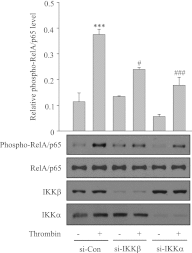
Effect of depletion of IKKα or IKKβ on thrombin-induced RelA/p65 phosphorylation. HPAECs were transfected with si-IKKα, siIKKβ, or si-Con for 24–36 hours and then challenged with thrombin (5 U/ml) for 1 hour. Total cell lysates were immunoblotted with an anti-phospho RelA/p65 (Ser536) antibody. Total RelA/p65 levels were used to monitor loading. Depletion of IKKα or IKKβ was determined by immunoblotting the lysates with an antibody to IKKα or IKKβ, respectively. The bar graph represents the effect of depletion of IKKα or IKKβ on thrombin-induced RelA/p65 phosphorylation normalized to total RelA/p65 level. Data are means (±SE) (n = 3–4 for each condition). ***P < 0.001 compared with untreated controls; ##P < 0.01 or #P < 0.05 compared with thrombin-treated control; #P < 0.05 compared with thrombin-treated control.

References

    1. Ghosh S, Hayden MS. New regulators of NF-kappa B in inflammation. Nat Rev Immunol 2008;8:837–848 - PubMed
    1. Rahman A, Fazal F. Blocking NF-kappaB: an inflammatory issue. Proc Am Thorac Soc 2011;8:497–503 - PMC - PubMed
    1. Bijli KM, Rahman ANF. NF-κB signaling in endothelium. In: Aird WC, editor. Endothelial biomedicine. New York: Cambridge University Press; 2007. pp. 784–795.
    1. Karin M. Nuclear factor-kappa B in cancer development and progression. Nature 2006;441:431–436 - PubMed
    1. Ye X, Ding J, Zhou X, Chen G, Liu SF. Divergent roles of endothelial NF-kappaB in multiple organ injury and bacterial clearance in mouse models of sepsis. J Exp Med 2008;205:1303–1315 - PMC - PubMed

Publication types

MeSH terms