. 2012 Nov;47(5):688-97.
doi: 10.1165/rcmb.2012-0161OC. Epub 2012 Jul 27.

Oxidative stress contributes to lung injury and barrier dysfunction via microtubule destabilization

Affiliations

Oxidative stress contributes to lung injury and barrier dysfunction via microtubule destabilization

Eric Kratzer et al. Am J Respir Cell Mol Biol. 2012 Nov.

Abstract

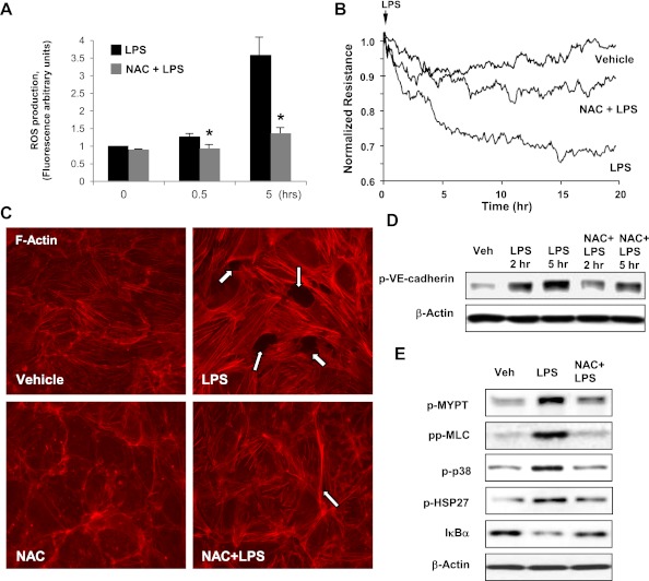
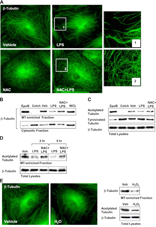
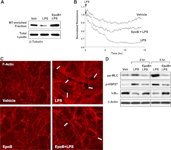
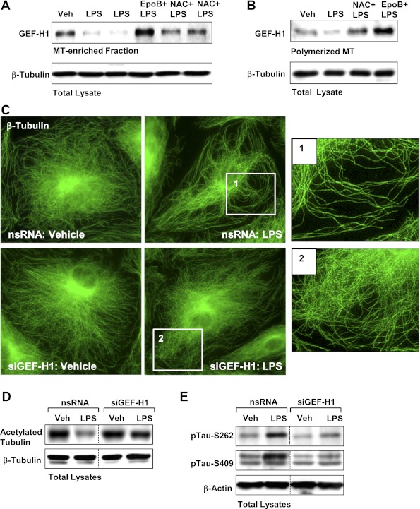
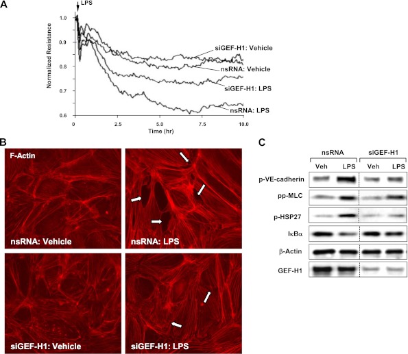
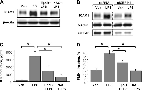
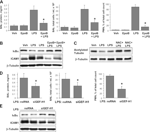
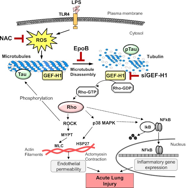
Oxidative stress is an important part of host innate immune response to foreign pathogens, such as bacterial LPS, but excessive activation of redox signaling may lead to pathologic endothelial cell (EC) activation and barrier dysfunction. Microtubules (MTs) play an important role in agonist-induced regulation of vascular endothelial permeability, but their impact in modulation of inflammation and EC barrier has not been yet investigated. This study examined the effects of LPS-induced oxidative stress on MT dynamics and the involvement of MTs in the LPS-induced mechanisms of Rho activation, EC permeability, and lung injury. LPS treatment of pulmonary vascular EC induced elevation of reactive oxygen species (ROS) and caused oxidative stress associated with EC hyperpermeability, cytoskeletal remodeling, and formation of paracellular gaps, as well as activation of Rho, p38 stress kinase, and NF-κB signaling, the hallmarks of endothelial barrier dysfunction. LPS also triggered ROS-dependent disassembly of the MT network, leading to activation of MT-dependent signaling. Stabilization of MTs with epothilone B, or inhibition of MT-associated guanine nucleotide exchange factor (GEF)-H1 activity by silencing RNA-mediated knockdown, suppressed LPS-induced EC barrier dysfunction in vitro, and attenuated vascular leak and lung inflammation in vivo. LPS disruptive effects were linked to activation of Rho signaling caused by LPS-induced MT disassembly and release of Rho-specific GEF-H1 from MTs. These studies demonstrate, for the first time, the mechanism of ROS-induced Rho activation via destabilization of MTs and GEF-H1-dependent activation of Rho signaling, leading to pulmonary EC barrier dysfunction and exacerbation of LPS-induced inflammation.


Figures

Figure 1.
Figure 1.
Role of oxidative stress in LPS-induced endothelial barrier disruption. Human pulmonary artery EC (HPAEC) monolayers were pretreated with vehicle or N-acetyl cysteine (NAC; 1 × 10⁻³ mol/L, 30 min) followed by LPS stimulation (300 ng/ml). (A) Reactive oxygen species (ROS) production was measured in live cells using fluorescent detection assay, as described in Materials and Methods. Data are expressed as means (±SD) of three independent experiments; *P < 0.05. (B) Measurements of transendothelial electrical resistance (TER) were monitored over 20 hours using an electrical cell-substrate impedance sensing system. (C) Analysis of actin cytoskeletal rearrangement was performed after 5 hours of LPS treatment by immunofluorescence staining with Texas Red phalloidin. Paracellular gaps are marked by arrows. (D and E) Phosphorylation of VE-cadherin (D), myosin-associated phosphatase type 1 (MYPT1), myosin light chain (MLC), p38, and heat shock protein (HSP) 27 (E) was determined by Western blot with corresponding phospho-specific antibodies. Degradation of IκBα was detected using pan IκBα antibodies. Equal protein loading was confirmed by determination of β-actin content in total cell lysates. Results are representative of three to five independent experiments.
Figure 2.
Figure 2.
Role of oxidative stress in LPS-induced microtubule (MT) remodeling. Human pulmonary artery endothelial cells (HPAECs) were pretreated with vehicle or NAC (1 × 10⁻³ mol/L, 30 min) followed by LPS (300 ng/ml). In control experiments, cells were treated with epothilone B (EpoB; 1 × 10⁻⁸ mol/L) or colchicine (Colch; 1 × 10⁻⁶ mol/L) for 30 minutes. (A) MT structure was analyzed by immunofluorescence staining for β-tubulin. Insets indicate high-magnification images of peripheral MT network. (B) Fractionation assay was performed and content of polymerized tubulin in MT-enriched fraction and depolymerized tubulin in cytosolic fraction was determined by Western blotting with β-tubulin antibodies. (C) Pools of acetylated MTs and tyrosinated MTs were determined in whole-cell lysates. Equal tubulin content was confirmed by probing of membranes for β-tubulin. (D) Effects of NAC on LPS-induced alteration of stable MTs were evaluated by Western blot analysis of MT-enriched fraction with antibodies against acetylated tubulin. Equal tubulin expression was confirmed by detection of β-tubulin in total cell lysates. (E) Effect of H2O2 (1 × 10⁻⁴ mol/L, 15 min) on MT structure was analyzed by immunofluorescence staining (upper panel), determination of polymerized tubulin content in MT-enriched fraction (lower left panel), and detection of acetylated tubulin in total cell lysates (lower right panel). Results are representative of three to six independent experiments.
Figure 3.
Figure 3.
Effect of MT stabilization on LPS-induced EC barrier dysfunction. HPAECs were pretreated with vehicle or epothilone B (1 × 10⁻⁸ mol/L, 30 min) followed by LPS (300 ng/ml) stimulation. (A) Content of polymerized tubulin in MT-enriched fraction was determined by Western blot analysis with β-tubulin antibodies. Equal tubulin expression was confirmed by detection of β-tubulin in total cell lysates. (B) TER measurements were performed over 15 hours. (C) Actin cytoskeletal remodeling after 5 hours of LPS treatment was examined by immunofluorescence staining with Texas Red–conjugated phalloidin. Paracellular gaps are marked by arrows. (D) Phosphorylation of MLC and HSP27, or IκBα content was analyzed by Western blotting. Equal protein loading was confirmed by determination of β-actin content in total cell lysates. Shown are representative results of three to five independent experiments.
Figure 4.
Figure 4.
Role of MT-associated guanine nucleotide exchange factor (GEF)-H1 in LPS-induced MT alteration. ECs were pretreated with vehicle, NAC (1 × 10⁻³ mol/L, 30 min), or epothilone B (1 × 10⁻⁸ mol/L, 30 min), followed by LPS (300 ng/ml) stimulation for 5 hours. (A and B) GEF-H1 content in MT-enriched fraction (A) or polymerized MT fraction (B) was determined by Western blot analysis with specific antibodies. Equal tubulin content was confirmed by detection of β-tubulin in total cell lysates. (CE) Human pulmonary ECs were transfected with GEF-H1–specific siRNA or nonspecific RNA followed by LPS stimulation. MT structure was analyzed by immunofluorescence staining for β-tubulin. Insets represent high-magnification images of peripheral MT network (C). Pool of stable MTs was determined by Western blot analysis with antibodies against acetylated tubulin. Equal tubulin content was confirmed by probing of membranes with β-tubulin antibodies (D). Tau phosphorylation after LPS challenge was analyzed by Western blot with specific antibodies. Equal protein loading was confirmed by determination of β-actin content in total cell lysates. Shown are representative results of three to four independent experiments.
Figure 5.
Figure 5.
Role of MT-associated GEF-H1 in LPS-induced MT alteration. HPAECs were transfected with GEF-H1–specific or nonspecific siRNA followed by LPS (300 ng/ml) stimulation. (A) TER measurements were performed over 10 hours. (B) Actin cytoskeletal remodeling after 5 hours of LPS treatment was examined by immunofluorescence staining with Texas Red–conjugated phalloidin. Paracellular gaps are marked by arrows. (C) Phosphorylation profile of proteins, IκBα, or GEF-H1 content was analyzed by Western blotting. Equal protein loading was confirmed by determination of β-actin content in total cell lysates. Shown are representative results of three to five independent experiments.
Figure 6.
Figure 6.
Role of MTs in LPS-induced endothelial activation. (A) ECs were pretreated with vehicle, NAC (1 × 10⁻³ mol/L, 30 min), or epothilone B (1 × 10⁻⁸ mol/L, 30 min) followed by LPS (300 ng/ml) stimulation for 5 hours. Intercellular adhesion molecule (ICAM)-1 expression was detected by Western blot with specific antibodies. β-actin staining was used as a normalization control. (B) HPAECs were transfected with GEF-H1–specific or nonspecific siRNA followed by LPS stimulation (300 ng/ml, 5 h). ICAM-1 expression was detected by Western blot analysis. Equal protein loading was confirmed by determination of β-actin content in total cell lysates. (C and D) Cells were pretreated with vehicle, NAC (1 × 10⁻³ mol/L, 30 min), or epothilone B (1 × 10⁻⁸ mol/L, 30 min) followed by LPS stimulation (20 ng/ml, 4 h). IL-8 production was determined in control and treated samples using an ELISA kit (C). Neutrophil migration assay was performed as described in Materials and Methods (D). Data are expressed as means (±SD) of five independent experiments; *P < 0.05.
Figure 7.
Figure 7.
Role of MTs in LPS-induced lung injury. (AC) C57BL/6J mice were challenged with LPS (0.63 mg/kg, intratracheally) with or without concurrent intravenous injection of epothilone B (4 × 10⁻⁶ mol/kg, intravenously) or NAC (2.5 × 10⁻³ mol/kg). Control animals were treated with sterile saline solution or epothilone B alone. (A) Protein concentration, total cell count, and neutrophil count were determined in bronchoalveolar lavage fluid collected 18 hours after treatments. Data are expressed as means (±SD) (n = 4–8 per condition); *P < 0.05, as compared with LPS treatment. (B) IκBα degradation and ICAM1 expression after LPS challenge with or without epothilone B treatment were determined in lung tissue homogenates by Western blot analysis with specific antibodies. Equal protein loading was confirmed by membrane reprobing with β-tubulin antibodies. (C) Levels of acetylated tubulin after LPS challenge with or without NAC treatment were determined in lung tissue homogenates by Western blot analysis with specific antibodies. Equal protein loading was confirmed by membrane reprobing with β-tubulin antibodies. (D and E) Mice were transfected with nonspecific or GEF-H1–specific siRNA for 72 hours followed by LPS (0.63 mg/kg, intratracheally) administration for 18 hours. (C) protein concentration, total cell count, and neutrophil count were determined in bronchoalveolar lavage fluid. Data are expressed as means (±SD) (n = 4–6 per condition; *P < 0.05). (D) IκBα degradation and ICAM1 expression after LPS challenge were determined in lung tissue homogenates from nonspecific or GEF-H1–specific siRNA-treated mice by Western blot analysis. Equal protein loading was confirmed by membrane reprobing with β-tubulin antibodies.
Figure 8.
Figure 8.
Proposed model of MT-dependent signaling in LPS-challenged lung endothelium (see explanation in the main text).

Similar articles

Cited by

References

    1. Mehta D, Malik AB. Signaling mechanisms regulating endothelial permeability. Physiol Rev 2006;86:279–367 - PubMed
    1. Lucas R, Verin AD, Black SM, Catravas JD. Regulators of endothelial and epithelial barrier integrity and function in acute lung injury. Biochem Pharmacol 2009;77:1763–1772 - PMC - PubMed
    1. Dosquet C, Weill D, Wautier JL. Molecular mechanism of blood monocyte adhesion to vascular endothelial cells. Nouv Rev Fr Hematol 1992;34:S55–S59 - PubMed
    1. Wang Q, Doerschuk CM. The signaling pathways induced by neutrophil–endothelial cell adhesion. Antioxid Redox Signal 2002;4:39–47 - PubMed
    1. Smith CW. Leukocyte–endothelial cell interactions. Semin Hematol 1993;30(4 Suppl 4):45–53; discussion 54–45. - PubMed

Publication types

MeSH terms